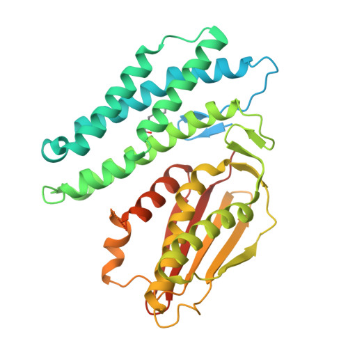
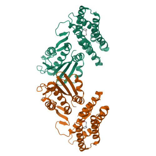
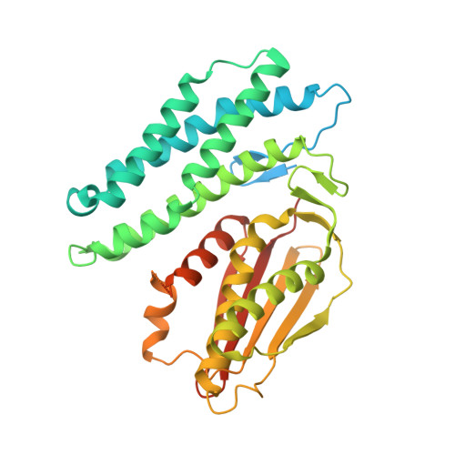
3TZ2

Crystal structure of branched-chain alpha-ketoacid dehydrogenase kinase/phenylbutyrate complex


Experimental Data Snapshot

  • Method: X-RAY DIFFRACTION
  • Resolution: 2.85 Å
  • R-Value Free: 0.242 
  • R-Value Work: 0.199 
  • R-Value Observed: 0.204 

wwPDB Validation   3D Report Full Report


Ligand Structure Quality Assessment 


This is version 1.3 of the entry. See complete history


Literature

Structure-based design and mechanisms of allosteric inhibitors for mitochondrial branched-chain alpha-ketoacid dehydrogenase kinase.

Tso, S.C.Qi, X.Gui, W.J.Chuang, J.L.Morlock, L.K.Wallace, A.L.Ahmed, K.Laxman, S.Campeau, P.M.Lee, B.H.Hutson, S.M.Tu, B.P.Williams, N.S.Tambar, U.K.Wynn, R.M.Chuang, D.T.

(2013) Proc Natl Acad Sci U S A 110: 9728-9733

  • DOI: https://doi.org/10.1073/pnas.1303220110
  • Primary Citation of Related Structures:  
    3TZ0, 3TZ2, 3TZ4, 3TZ5, 4DZY, 4H7Q, 4H81, 4H85

  • PubMed Abstract: 

    The branched-chain amino acids (BCAAs) leucine, isoleucine, and valine are elevated in maple syrup urine disease, heart failure, obesity, and type 2 diabetes. BCAA homeostasis is controlled by the mitochondrial branched-chain α-ketoacid dehydrogenase complex (BCKDC), which is negatively regulated by the specific BCKD kinase (BDK). Here, we used structure-based design to develop a BDK inhibitor, (S)-α-chloro-phenylpropionic acid [(S)-CPP]. Crystal structures of the BDK-(S)-CPP complex show that (S)-CPP binds to a unique allosteric site in the N-terminal domain, triggering helix movements in BDK. These conformational changes are communicated to the lipoyl-binding pocket, which nullifies BDK activity by blocking its binding to the BCKDC core. Administration of (S)-CPP to mice leads to the full activation and dephosphorylation of BCKDC with significant reduction in plasma BCAA concentrations. The results buttress the concept of targeting mitochondrial BDK as a pharmacological approach to mitigate BCAA accumulation in metabolic diseases and heart failure.


  • Organizational Affiliation

    Department of Biochemistry, University of Texas Southwestern Medical Center, Dallas, TX 75390, USA.


Macromolecules
Find similar proteins by:  (by identity cutoff)  |  3D Structure
Entity ID: 1
MoleculeChains Sequence LengthOrganismDetailsImage
[3-methyl-2-oxobutanoate dehydrogenase [lipoamide]] kinase, mitochondrial418Rattus norvegicusMutation(s): 0 
Gene Names: Bckdk
EC: 2.7.11.4 (PDB Primary Data), 2.7.11.1 (UniProt)
UniProt
Find proteins for Q00972 (Rattus norvegicus)
Explore Q00972 
Go to UniProtKB:  Q00972
Entity Groups  
Sequence Clusters30% Identity50% Identity70% Identity90% Identity95% Identity100% Identity
UniProt GroupQ00972
Sequence Annotations
Expand
  • Reference Sequence
Small Molecules
Ligands 1 Unique
IDChains Name / Formula / InChI Key2D Diagram3D Interactions
CLT
Query on CLT

Download Ideal Coordinates CCD File 
B [auth A]4-PHENYL-BUTANOIC ACID
C10 H12 O2
OBKXEAXTFZPCHS-UHFFFAOYSA-N
Binding Affinity Annotations 
IDSourceBinding Affinity
CLT PDBBind:  3TZ2 Kd: 5700 (nM) from 1 assay(s)
Experimental Data & Validation

Experimental Data

  • Method: X-RAY DIFFRACTION
  • Resolution: 2.85 Å
  • R-Value Free: 0.242 
  • R-Value Work: 0.199 
  • R-Value Observed: 0.204 
  • Space Group: P 42 21 2
Unit Cell:
Length ( Å )Angle ( ˚ )
a = 128.201α = 90
b = 128.201β = 90
c = 72.111γ = 90
Software Package:
Software NamePurpose
HKL-2000data collection
PHENIXmodel building
PHENIXrefinement
HKL-2000data reduction
HKL-2000data scaling
PHENIXphasing

Structure Validation

View Full Validation Report


Ligand Structure Quality Assessment 


Entry History 

Deposition Data

Revision History  (Full details and data files)

  • Version 1.0: 2012-10-03
    Type: Initial release
  • Version 1.1: 2013-05-29
    Changes: Database references
  • Version 1.2: 2019-09-18
    Changes: Data collection, Database references
  • Version 1.3: 2024-02-28
    Changes: Data collection, Database references, Derived calculations