3W6G

Structure of peroxiredoxin from anaerobic hyperthermophilic archaeon Pyrococcus horikoshii


Experimental Data Snapshot

  • Method: X-RAY DIFFRACTION
  • Resolution: 2.25 Å
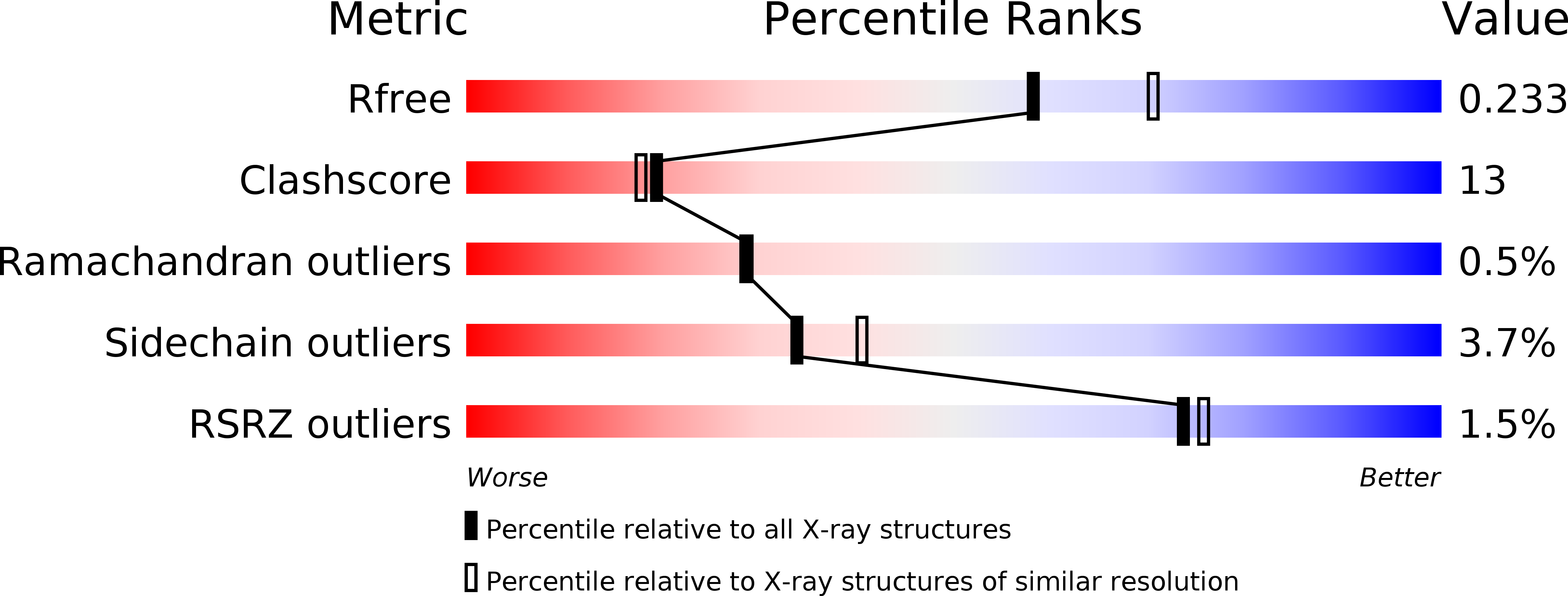
  • R-Value Free: 0.237 
  • R-Value Work: 0.198 
  • R-Value Observed: 0.198 

wwPDB Validation   3D Report Full Report


This is version 1.2 of the entry. See complete history


Literature

Structure of peroxiredoxin from the anaerobic hyperthermophilic archaeon Pyrococcus horikoshii

Nakamura, T.Mori, A.Niiyama, M.Matsumura, H.Tokuyama, C.Morita, J.Uegaki, K.Inoue, T.

(2013) Acta Crystallogr Sect F Struct Biol Cryst Commun 69: 719-722

  • DOI: https://doi.org/10.1107/S1744309113014036
  • Primary Citation of Related Structures:  
    3W6G

  • PubMed Abstract: 

    The crystal structure of peroxiredoxin from the anaerobic hyperthermophilic archaeon Pyrococcus horikoshii (PhPrx) was determined at a resolution of 2.25 Å. The overall structure was a ring-type decamer consisting of five homodimers. Citrate, which was included in the crystallization conditions, was bound to the peroxidatic cysteine of the active site, with two O atoms of the carboxyl group mimicking those of the substrate hydrogen peroxide. PhPrx lacked the C-terminal tail that forms a 32-residue extension of the protein in the homologous peroxiredoxin from Aeropyrum pernix (ApPrx).


  • Organizational Affiliation

    National Institute of Advanced Industrial Science and Technology, Ikeda, Osaka 563-8577, Japan. nakamura-t@aist.go.jp


Macromolecules
Find similar proteins by:  (by identity cutoff)  |  3D Structure
Entity ID: 1
MoleculeChains Sequence LengthOrganismDetailsImage
Probable peroxiredoxin
A, B, C, D, E
A, B, C, D, E, F, G, H, I, J, K, L, M, N, O, P, Q, R, S, T
216Pyrococcus horikoshii OT3Mutation(s): 0 
Gene Names: PH1217
EC: 1.11.1.15 (PDB Primary Data), 1.11.1.24 (UniProt)
UniProt
Find proteins for O58966 (Pyrococcus horikoshii (strain ATCC 700860 / DSM 12428 / JCM 9974 / NBRC 100139 / OT-3))
Explore O58966 
Go to UniProtKB:  O58966
Entity Groups  
Sequence Clusters30% Identity50% Identity70% Identity90% Identity95% Identity100% Identity
UniProt GroupO58966
Sequence Annotations
Expand
  • Reference Sequence
Small Molecules
Ligands 1 Unique
IDChains Name / Formula / InChI Key2D Diagram3D Interactions
FLC
Query on FLC

Download Ideal Coordinates CCD File 
AA [auth G]
BA [auth H]
CA [auth I]
DA [auth J]
EA [auth K]
AA [auth G],
BA [auth H],
CA [auth I],
DA [auth J],
EA [auth K],
FA [auth L],
GA [auth M],
HA [auth N],
IA [auth O],
JA [auth P],
KA [auth Q],
LA [auth R],
MA [auth S],
NA [auth T],
U [auth A],
V [auth B],
W [auth C],
X [auth D],
Y [auth E],
Z [auth F]
CITRATE ANION
C6 H5 O7
KRKNYBCHXYNGOX-UHFFFAOYSA-K
Experimental Data & Validation

Experimental Data

  • Method: X-RAY DIFFRACTION
  • Resolution: 2.25 Å
  • R-Value Free: 0.237 
  • R-Value Work: 0.198 
  • R-Value Observed: 0.198 
  • Space Group: P 1 21 1
Unit Cell:
Length ( Å )Angle ( ˚ )
a = 125.354α = 90
b = 94.733β = 103.31
c = 228.204γ = 90
Software Package:
Software NamePurpose
HKL-2000data collection
MOLREPphasing
CNSrefinement
HKL-2000data reduction
HKL-2000data scaling

Structure Validation

View Full Validation Report


Entry History 

Deposition Data

Revision History  (Full details and data files)

  • Version 1.0: 2013-07-10
    Type: Initial release
  • Version 1.1: 2013-12-11
    Changes: Database references
  • Version 1.2: 2024-03-20
    Changes: Data collection, Database references, Derived calculations