3C7M

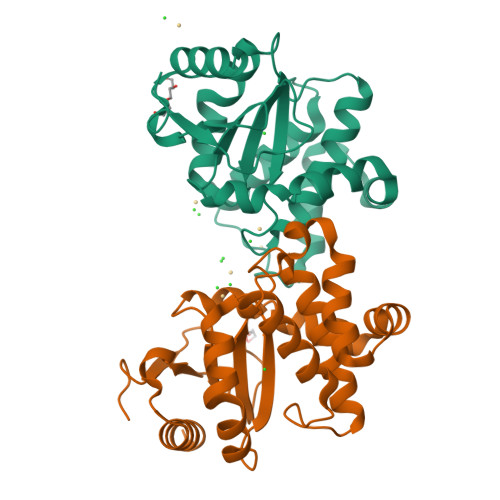
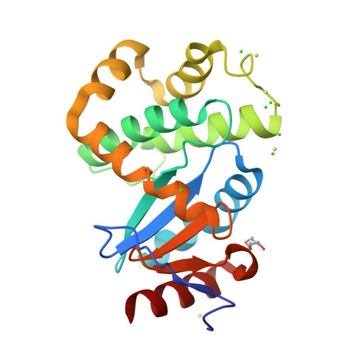
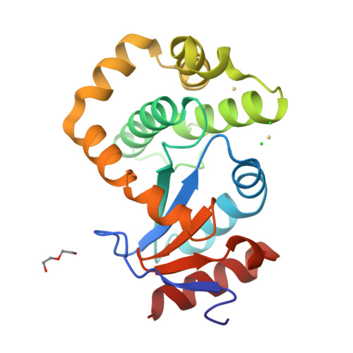
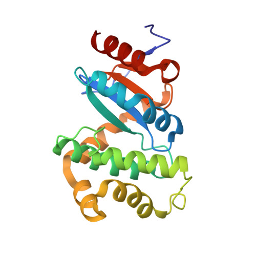
Crystal structure of reduced DsbL


Experimental Data Snapshot

  • Method: X-RAY DIFFRACTION
  • Resolution: 1.55 Å
  • R-Value Free: 0.189 
  • R-Value Work: 0.164 
  • R-Value Observed: 0.165 

Starting Model: experimental
View more details

wwPDB Validation   3D Report Full Report


This is version 1.3 of the entry. See complete history


Literature

DsbL and DsbI form a specific dithiol oxidase system for periplasmic arylsulfate sulfotransferase in uropathogenic Escherichia coli.

Grimshaw, J.P.Stirnimann, C.U.Brozzo, M.S.Malojcic, G.Grutter, M.G.Capitani, G.Glockshuber, R.

(2008) J Mol Biol 380: 667-680

  • DOI: https://doi.org/10.1016/j.jmb.2008.05.031
  • Primary Citation of Related Structures:  
    3C7M

  • PubMed Abstract: 

    Disulfide bond formation in the Escherichia coli periplasm requires the transfer of electrons from substrate proteins to DsbA, which is recycled as an oxidant by the membrane protein DsbB. The highly virulent, uropathogenic E. coli strain CFT073 contains a second, homologous pair of proteins, DsbL and DsbI, which are encoded in a tri-cistronic operon together with a periplasmic, uropathogen-specific arylsulfate sulfotransferase (ASST). We show that DsbL and DsbI form a functional redox pair, and that ASST is a substrate of DsbL/DsbI in vivo. DsbL is the most reactive oxidizing thioredoxin-like protein known to date. In contrast to DsbA, however, DsbL oxidizes reduced RNaseA with a much lower rate and prevents unspecific aggregation of reduced insulin. The 1.55 A resolution crystal structure of reduced DsbL provides insight into the reduced state of thioredoxin-like dithiol oxidases at high resolution, and reveals an unusual cluster of basic residues stabilizing the thiolate anion of the nucleophilic active-site cysteine. We propose that the DsbL/DsbI pair of uropathogenic E. coli was acquired as an additional, specific redox couple that guarantees biological activity of ASST.


  • Organizational Affiliation

    Institut für Molekularbiologie und Biophysik, ETH Zürich, CH-8093 Zürich, Switzerland.


Macromolecules
Find similar proteins by:  (by identity cutoff)  |  3D Structure
Entity ID: 1
MoleculeChains Sequence LengthOrganismDetailsImage
Thiol:disulfide interchange protein dsbA-like
A, B
195Escherichia coliMutation(s): 0 
UniProt
Find proteins for P0A4L7 (Escherichia coli O6:H1 (strain CFT073 / ATCC 700928 / UPEC))
Explore P0A4L7 
Go to UniProtKB:  P0A4L7
Entity Groups  
Sequence Clusters30% Identity50% Identity70% Identity90% Identity95% Identity100% Identity
UniProt GroupP0A4L7
Sequence Annotations
Expand
  • Reference Sequence
Small Molecules
Ligands 4 Unique
IDChains Name / Formula / InChI Key2D Diagram3D Interactions
PGE
Query on PGE

Download Ideal Coordinates CCD File 
N [auth A]TRIETHYLENE GLYCOL
C6 H14 O4
ZIBGPFATKBEMQZ-UHFFFAOYSA-N
CD
Query on CD

Download Ideal Coordinates CCD File 
C [auth A]
D [auth A]
E [auth A]
F [auth A]
O [auth B]
C [auth A],
D [auth A],
E [auth A],
F [auth A],
O [auth B],
P [auth B]
CADMIUM ION
Cd
WLZRMCYVCSSEQC-UHFFFAOYSA-N
PEG
Query on PEG

Download Ideal Coordinates CCD File 
T [auth B]DI(HYDROXYETHYL)ETHER
C4 H10 O3
MTHSVFCYNBDYFN-UHFFFAOYSA-N
CL
Query on CL

Download Ideal Coordinates CCD File 
G [auth A]
H [auth A]
I [auth A]
J [auth A]
K [auth A]
G [auth A],
H [auth A],
I [auth A],
J [auth A],
K [auth A],
L [auth A],
M [auth A],
Q [auth B],
R [auth B],
S [auth B]
CHLORIDE ION
Cl
VEXZGXHMUGYJMC-UHFFFAOYSA-M
Experimental Data & Validation

Experimental Data

  • Method: X-RAY DIFFRACTION
  • Resolution: 1.55 Å
  • R-Value Free: 0.189 
  • R-Value Work: 0.164 
  • R-Value Observed: 0.165 
  • Space Group: P 1 21 1
Unit Cell:
Length ( Å )Angle ( ˚ )
a = 50.945α = 90
b = 66.634β = 107.02
c = 77.891γ = 90
Software Package:
Software NamePurpose
REFMACrefinement
XDSdata reduction
XSCALEdata scaling
PHASERphasing

Structure Validation

View Full Validation Report


Entry History 

Deposition Data

Revision History  (Full details and data files)

  • Version 1.0: 2008-07-15
    Type: Initial release
  • Version 1.1: 2011-07-13
    Changes: Version format compliance
  • Version 1.2: 2024-02-21
    Changes: Data collection, Database references, Derived calculations
  • Version 1.3: 2024-04-03
    Changes: Refinement description