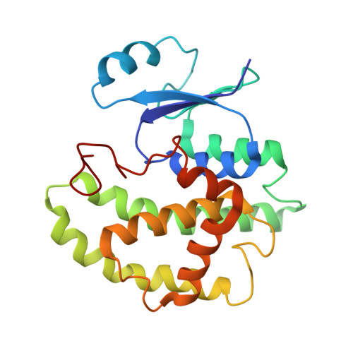
Using affinity chromatography to engineer and characterize pH-dependent protein switches.
Sagermann, M., Chapleau, R.R., DeLorimier, E., Lei, M.(2009) Protein Sci 18: 217-228
- PubMed: 19177365
- DOI: https://doi.org/10.1002/pro.23
- Primary Citation of Related Structures:
3CRT, 3CRU, 3D0Z - PubMed Abstract:
Conformational changes play important roles in the regulation of many enzymatic reactions. Specific motions of side chains, secondary structures, or entire protein domains facilitate the precise control of substrate selection, binding, and catalysis. Likewise, the engineering of allostery into proteins is envisioned to enable unprecedented control of chemical reactions and molecular assembly processes. We here study the structural effects of engineered ionizable residues in the core of the glutathione-S-transferase to convert this protein into a pH-dependent allosteric protein. The underlying rational of these substitutions is that in the neutral state, an uncharged residue is compatible with the hydrophobic environment. In the charged state, however, the residue will invoke unfavorable interactions, which are likely to induce conformational changes that will affect the function of the enzyme. To test this hypothesis, we have engineered a single aspartate, cysteine, or histidine residue at a distance from the active site into the protein. All of the mutations exhibit a dramatic effect on the protein's affinity to bind glutathione. Whereas the aspartate or histidine mutations result in permanently nonbinding or binding versions of the protein, respectively, mutant GST50C exhibits distinct pH-dependent GSH-binding affinity. The crystal structures of the mutant protein GST50C under ionizing and nonionizing conditions reveal the recruitment of water molecules into the hydrophobic core to produce conformational changes that influence the protein's active site. The methodology described here to create and characterize engineered allosteric proteins through affinity chromatography may lead to a general approach to engineer effector-specific allostery into a protein structure.
Organizational Affiliation:
Department of Chemistry and Biochemistry, Interdepartmental Program in BioMolecular Science and Engineering, University of California, Santa Barbara, California 93106-9510, USA. sagermann@chem.ucsb.edu


