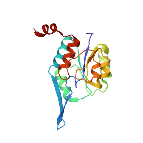
3E84

Structure-function Analysis of 2-Keto-3-deoxy-D-glycero-D-galacto-nononate-9-phosphate (KDN) Phosphatase Defines a New Clad Within the Type C0 HAD Subfamily


Experimental Data Snapshot

  • Method: X-RAY DIFFRACTION
  • Resolution: 1.85 Å
  • R-Value Free: 0.218 
  • R-Value Work: 0.176 
  • R-Value Observed: 0.178 

Starting Model: experimental
View more details

wwPDB Validation   3D Report Full Report


This is version 1.5 of the entry. See complete history


Literature

Structure-Function Analysis of 2-Keto-3-deoxy-D-glycero-D-galactonononate-9-phosphate Phosphatase Defines Specificity Elements in Type C0 Haloalkanoate Dehalogenase Family Members.

Lu, Z.Wang, L.Dunaway-Mariano, D.Allen, K.N.

(2009) J Biol Chem 284: 1224-1233

  • DOI: https://doi.org/10.1074/jbc.M807056200
  • Primary Citation of Related Structures:  
    3E81, 3E84, 3E8M

  • PubMed Abstract: 

    The phosphotransferases of the haloalkanoate dehalogenase superfamily (HADSF) act upon a wide range of metabolites in all eukaryotes and prokaryotes and thus constitute a significant force in cell function. The challenge posed for biochemical function assignment of HADSF members is the identification of the structural determinants that target a specific metabolite. The "8KDOP" subfamily of the HADSF is defined by the known structure and catalytic activity of 2-keto-3-deoxy-8-phospho-d-manno-octulosonic acid (KDO-8-P) phosphatase. Homologues of this enzyme have been uniformly annotated as KDO-8-P phosphatase. One such gene, BT1713, from the Bacteroides thetaiotaomicron genome was recently found to encode the enzyme 2-keto-3-deoxy-d-glycero-d-galacto-9-phosphonononic acid (KDN-9-P) phosphatase in the biosynthetic pathway of the 9-carbon alpha-keto acid, 2-keto-3-deoxy-d-glycero-d-galactonononic acid (KDN). To find the structural elements that provide substrate-specific interactions and to allow identification of genomic sequence markers, the x-ray crystal structures of BT1713 liganded to the cofactor Mg(2+)and complexed with tungstate or VO(3)(-)/Neu5Ac were determined to 1.1, 1.85, and 1.63 A resolution, respectively. The structures define the active site to be at the subunit interface and, as confirmed by steady-state kinetics and site-directed mutagenesis, reveal Arg-64(*), Lys-67(*), and Glu-56 to be the key residues involved in sugar binding that are essential for BT1713 catalytic function. Bioinformatic analyses of the differentially conserved residues between BT1713 and KDO-8-P phosphatase homologues guided by the knowledge of the structure-based specificity determinants define Glu-56 and Lys-67(*) to be the key residues that can be used in future annotations.


  • Organizational Affiliation

    Department of Chemistry, Boston University, Boston, Massachusetts 02215-2521, USA.


Macromolecules
Find similar proteins by:  (by identity cutoff)  |  3D Structure
Entity ID: 1
MoleculeChains Sequence LengthOrganismDetailsImage
Acylneuraminate cytidylyltransferase
A, B, C, D
164Bacteroides thetaiotaomicronMutation(s): 0 
Gene Names: BT1713
EC: 3.1.3.103
UniProt
Find proteins for Q8A712 (Bacteroides thetaiotaomicron (strain ATCC 29148 / DSM 2079 / JCM 5827 / CCUG 10774 / NCTC 10582 / VPI-5482 / E50))
Explore Q8A712 
Go to UniProtKB:  Q8A712
Entity Groups  
Sequence Clusters30% Identity50% Identity70% Identity90% Identity95% Identity100% Identity
UniProt GroupQ8A712
Sequence Annotations
Expand
  • Reference Sequence
Small Molecules
Ligands 4 Unique
IDChains Name / Formula / InChI Key2D Diagram3D Interactions
WO4
Query on WO4

Download Ideal Coordinates CCD File 
F [auth A],
K [auth B],
N [auth C],
Q [auth D]
TUNGSTATE(VI)ION
O4 W
PBYZMCDFOULPGH-UHFFFAOYSA-N
PEG
Query on PEG

Download Ideal Coordinates CCD File 
G [auth A],
H [auth A],
L [auth B],
O [auth C]
DI(HYDROXYETHYL)ETHER
C4 H10 O3
MTHSVFCYNBDYFN-UHFFFAOYSA-N
EDO
Query on EDO

Download Ideal Coordinates CCD File 
I [auth A]1,2-ETHANEDIOL
C2 H6 O2
LYCAIKOWRPUZTN-UHFFFAOYSA-N
MG
Query on MG

Download Ideal Coordinates CCD File 
E [auth A],
J [auth B],
M [auth C],
P [auth D]
MAGNESIUM ION
Mg
JLVVSXFLKOJNIY-UHFFFAOYSA-N
Experimental Data & Validation

Experimental Data

  • Method: X-RAY DIFFRACTION
  • Resolution: 1.85 Å
  • R-Value Free: 0.218 
  • R-Value Work: 0.176 
  • R-Value Observed: 0.178 
  • Space Group: P 21 21 2
Unit Cell:
Length ( Å )Angle ( ˚ )
a = 82.086α = 90
b = 107.336β = 90
c = 75.31γ = 90
Software Package:
Software NamePurpose
DENZOdata reduction
SCALEPACKdata scaling
PHENIXrefinement
PDB_EXTRACTdata extraction
CrystalCleardata collection
MOLREPphasing

Structure Validation

View Full Validation Report


Entry History 

Deposition Data

Revision History  (Full details and data files)

  • Version 1.0: 2008-11-04
    Type: Initial release
  • Version 1.1: 2011-07-13
    Changes: Version format compliance
  • Version 1.2: 2011-08-31
    Changes: Refinement description
  • Version 1.3: 2011-11-16
    Changes: Atomic model
  • Version 1.4: 2017-10-25
    Changes: Refinement description
  • Version 1.5: 2023-08-30
    Changes: Data collection, Database references, Derived calculations, Refinement description