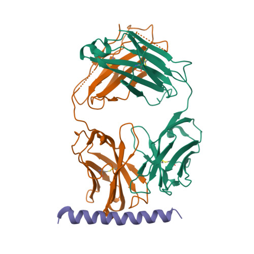
3EFD

The crystal structure of the cytoplasmic domain of KcsA


Experimental Data Snapshot

  • Method: X-RAY DIFFRACTION
  • Resolution: 2.60 Å
  • R-Value Free: 0.276 
  • R-Value Work: 0.225 
  • R-Value Observed: 0.230 

wwPDB Validation   3D Report Full Report


This is version 1.1 of the entry. See complete history


Literature

Crystal structure of full-length KcsA in its closed conformation.

Uysal, S.Vasquez, V.Tereshko, V.Esaki, K.Fellouse, F.A.Sidhu, S.S.Koide, S.Perozo, E.Kossiakoff, A.

(2009) Proc Natl Acad Sci U S A 106: 6644-6649

  • DOI: https://doi.org/10.1073/pnas.0810663106
  • Primary Citation of Related Structures:  
    3EFD, 3EFF

  • PubMed Abstract: 

    KcsA is a proton-activated, voltage-modulated K(+) channel that has served as the archetype pore domain in the Kv channel superfamily. Here, we have used synthetic antigen-binding fragments (Fabs) as crystallographic chaperones to determine the structure of full-length KcsA at 3.8 A, as well as that of its isolated C-terminal domain at 2.6 A. The structure of the full-length KcsA-Fab complex reveals a well-defined, 4-helix bundle that projects approximately 70 A toward the cytoplasm. This bundle promotes a approximately 15 degree bending in the inner bundle gate, tightening its diameter and shifting the narrowest point 2 turns of helix below. Functional analysis of the full-length KcsA-Fab complex suggests that the C-terminal bundle remains whole during gating. We suggest that this structure likely represents the physiologically relevant closed conformation of KcsA.


  • Organizational Affiliation

    Department of Biochemistry and Molecular Biology, and Institute for Biophysical Dynamics, University of Chicago, Chicago, IL 60637, USA.


Macromolecules
Find similar proteins by:  (by identity cutoff)  |  3D Structure
Entity ID: 1
MoleculeChains Sequence LengthOrganismDetailsImage
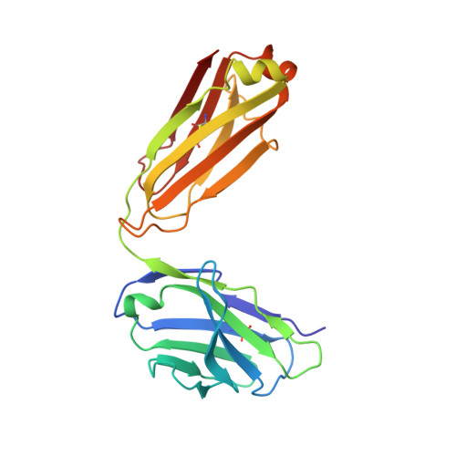
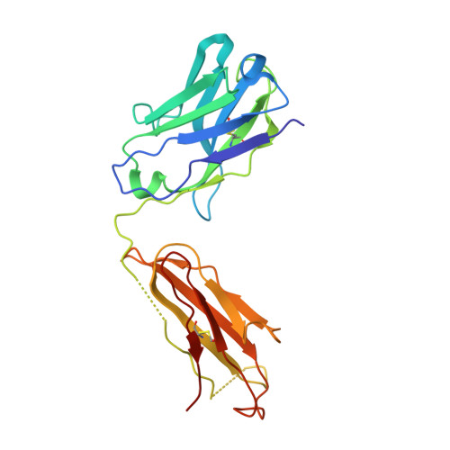
FabLA [auth L]211Mus musculusMutation(s): 0 
Sequence Annotations
Expand
  • Reference Sequence
Find similar proteins by:  (by identity cutoff)  |  3D Structure
Entity ID: 2
MoleculeChains Sequence LengthOrganismDetailsImage
FabHB [auth H]222Mus musculusMutation(s): 0 
Sequence Annotations
Expand
  • Reference Sequence
Find similar proteins by:  (by identity cutoff)  |  3D Structure
Entity ID: 3
MoleculeChains Sequence LengthOrganismDetailsImage
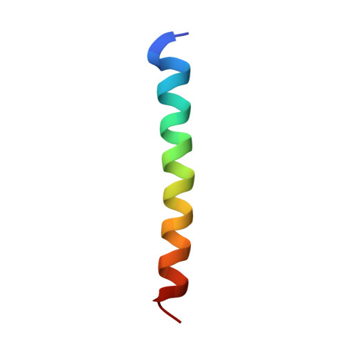
KcsAC [auth K]30Escherichia coliMutation(s): 0 
Sequence Annotations
Expand
  • Reference Sequence
Experimental Data & Validation

Experimental Data

  • Method: X-RAY DIFFRACTION
  • Resolution: 2.60 Å
  • R-Value Free: 0.276 
  • R-Value Work: 0.225 
  • R-Value Observed: 0.230 
  • Space Group: I 4
Unit Cell:
Length ( Å )Angle ( ˚ )
a = 115.499α = 90
b = 115.499β = 90
c = 76.816γ = 90
Software Package:
Software NamePurpose
ADSCdata collection
CCP4model building
REFMACrefinement
HKL-2000data reduction
HKL-2000data scaling
CCP4phasing

Structure Validation

View Full Validation Report


Entry History 

Deposition Data

Revision History  (Full details and data files)

  • Version 1.0: 2009-04-14
    Type: Initial release
  • Version 1.1: 2011-07-13
    Changes: Advisory, Refinement description, Version format compliance