3HBL

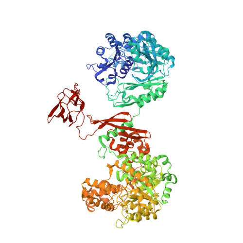
Crystal Structure of S. aureus Pyruvate Carboxylase T908A Mutant


Experimental Data Snapshot

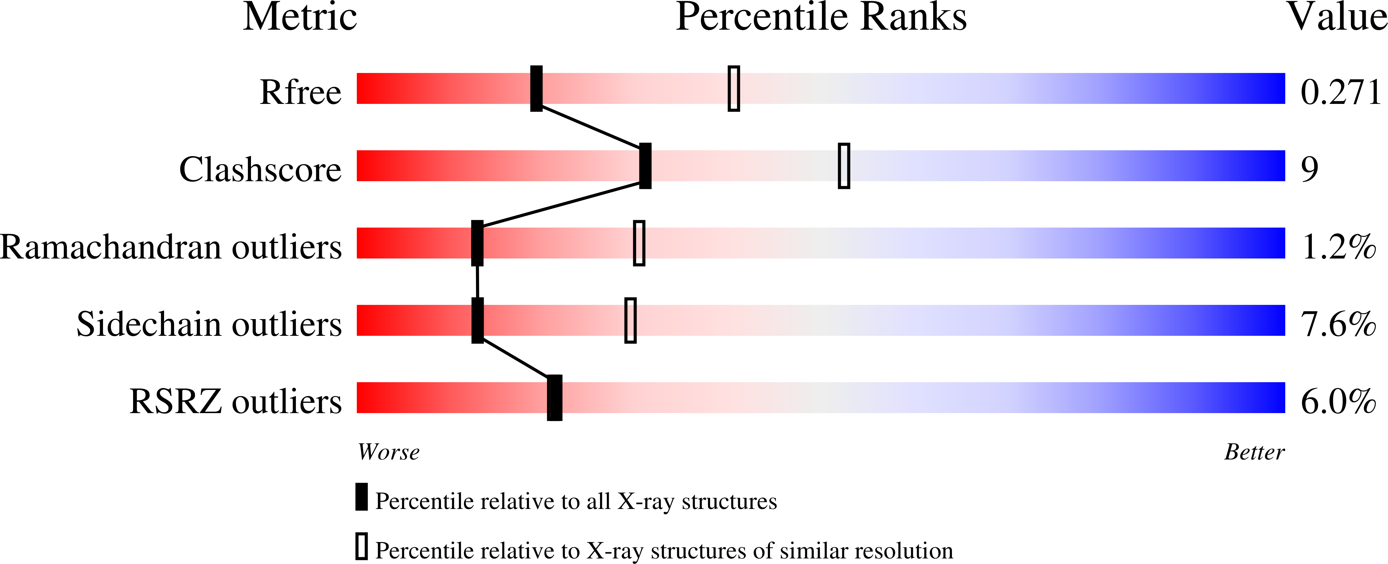
  • Method: X-RAY DIFFRACTION
  • Resolution: 2.71 Å
  • R-Value Free: 0.279 
  • R-Value Work: 0.228 
  • R-Value Observed: 0.230 

wwPDB Validation   3D Report Full Report


Ligand Structure Quality Assessment 


This is version 1.3 of the entry. See complete history


Literature

A Symmetrical Tetramer for S. aureus Pyruvate Carboxylase in Complex with Coenzyme A.

Yu, L.P.Xiang, S.Lasso, G.Gil, D.Valle, M.Tong, L.

(2009) Structure 17: 823-832

  • DOI: https://doi.org/10.1016/j.str.2009.04.008
  • Primary Citation of Related Structures:  
    3HB9, 3HBL, 3HO8

  • PubMed Abstract: 

    Pyruvate carboxylase (PC) is a conserved metabolic enzyme with important cellular functions. We report crystallographic and cryo-electron microscopy (EM) studies of Staphylococcus aureus PC (SaPC) in complex with acetyl-CoA, an allosteric activator, and mutagenesis, biochemical, and structural studies of the biotin binding site of its carboxyltransferase (CT) domain. The disease-causing A610T mutation abolishes catalytic activity by blocking biotin binding to the CT active site, and Thr908 might play a catalytic role in the CT reaction. The crystal structure of SaPC in complex with CoA reveals a symmetrical tetramer, with one CoA molecule bound to each monomer, and cryo-EM studies confirm the symmetrical nature of the tetramer. These observations are in sharp contrast to the highly asymmetrical tetramer of Rhizobium etli PC in complex with ethyl-CoA. Our structural information suggests that acetyl-CoA promotes a conformation for the dimer of the biotin carboxylase domain of PC that might be catalytically more competent.


  • Organizational Affiliation

    Department of Biological Sciences, Columbia University, New York, NY 10027, USA.


Macromolecules
Find similar proteins by:  (by identity cutoff)  |  3D Structure
Entity ID: 1
MoleculeChains Sequence LengthOrganismDetailsImage
Pyruvate carboxylase
A, B, C, D
1,150Staphylococcus aureus subsp. aureus Mu50Mutation(s): 1 
Gene Names: pycAPyruvate CarboxylaseSAV1114
EC: 6.4.1.1
UniProt
Find proteins for A0A0H3JRU9 (Staphylococcus aureus (strain Mu50 / ATCC 700699))
Explore A0A0H3JRU9 
Go to UniProtKB:  A0A0H3JRU9
Entity Groups  
Sequence Clusters30% Identity50% Identity70% Identity90% Identity95% Identity100% Identity
UniProt GroupA0A0H3JRU9
Sequence Annotations
Expand
  • Reference Sequence
Small Molecules
Ligands 3 Unique
IDChains Name / Formula / InChI Key2D Diagram3D Interactions
ADP
Query on ADP

Download Ideal Coordinates CCD File 
J [auth C]ADENOSINE-5'-DIPHOSPHATE
C10 H15 N5 O10 P2
XTWYTFMLZFPYCI-KQYNXXCUSA-N
BTI
Query on BTI

Download Ideal Coordinates CCD File 
E [auth A],
G [auth B],
I [auth C],
L [auth D]
5-(HEXAHYDRO-2-OXO-1H-THIENO[3,4-D]IMIDAZOL-6-YL)PENTANAL
C10 H16 N2 O2 S
ARDNWGMSCXSPBF-CIUDSAMLSA-N
MN
Query on MN

Download Ideal Coordinates CCD File 
F [auth A],
H [auth B],
K [auth C],
M [auth D]
MANGANESE (II) ION
Mn
WAEMQWOKJMHJLA-UHFFFAOYSA-N
Experimental Data & Validation

Experimental Data

  • Method: X-RAY DIFFRACTION
  • Resolution: 2.71 Å
  • R-Value Free: 0.279 
  • R-Value Work: 0.228 
  • R-Value Observed: 0.230 
  • Space Group: P 1 21 1
Unit Cell:
Length ( Å )Angle ( ˚ )
a = 96.532α = 90
b = 257.154β = 114.35
c = 130.318γ = 90
Software Package:
Software NamePurpose
DENZOdata reduction
SCALEPACKdata scaling
COMOphasing
REFMACrefinement
PDB_EXTRACTdata extraction

Structure Validation

View Full Validation Report


Ligand Structure Quality Assessment 


Entry History 

Deposition Data

Revision History  (Full details and data files)

  • Version 1.0: 2009-06-30
    Type: Initial release
  • Version 1.1: 2011-07-13
    Changes: Version format compliance
  • Version 1.2: 2017-11-01
    Changes: Refinement description
  • Version 1.3: 2021-10-13
    Changes: Database references, Derived calculations