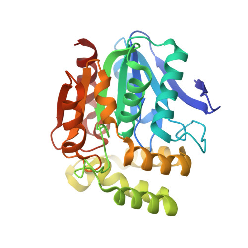
3HEA

The L29P/L124I mutation of Pseudomonas fluorescens esterase


Experimental Data Snapshot

  • Method: X-RAY DIFFRACTION
  • Resolution: 1.90 Å
  • R-Value Free: 0.212 
  • R-Value Work: 0.190 
  • R-Value Observed: 0.191 

Starting Model: experimental
View more details

wwPDB Validation   3D Report Full Report


This is version 1.4 of the entry. See complete history


Literature

Switching catalysis from hydrolysis to perhydrolysis in Pseudomonas fluorescens esterase.

Yin de, L.T.Bernhardt, P.Morley, K.L.Jiang, Y.Cheeseman, J.D.Purpero, V.Schrag, J.D.Kazlauskas, R.J.

(2010) Biochemistry 49: 1931-1942

  • DOI: https://doi.org/10.1021/bi9021268
  • Primary Citation of Related Structures:  
    3HEA, 3HI4

  • PubMed Abstract: 

    Many serine hydrolases catalyze perhydrolysis, the reversible formation of peracids from carboxylic acids and hydrogen peroxide. Recently, we showed that a single amino acid substitution in the alcohol binding pocket, L29P, in Pseudomonas fluorescens (SIK WI) aryl esterase (PFE) increased the specificity constant of PFE for peracetic acid formation >100-fold [Bernhardt et al. (2005) Angew. Chem., Int. Ed. 44, 2742]. In this paper, we extend this work to address the three following questions. First, what is the molecular basis of the increase in perhydrolysis activity? We previously proposed that the L29P substitution creates a hydrogen bond between the enzyme and hydrogen peroxide in the transition state. Here we report two X-ray structures of L29P PFE that support this proposal. Both structures show a main chain carbonyl oxygen closer to the active site serine as expected. One structure further shows acetate in the active site in an orientation consistent with reaction by an acyl-enzyme mechanism. We also detected an acyl-enzyme intermediate in the hydrolysis of epsilon-caprolactone by mass spectrometry. Second, can we further increase perhydrolysis activity? We discovered that the reverse reaction, hydrolysis of peracetic acid to acetic acid and hydrogen peroxide, occurs at nearly the diffusion limited rate. Since the reverse reaction cannot increase further, neither can the forward reaction. Consistent with this prediction, two variants with additional amino acid substitutions showed 2-fold higher k(cat), but K(m) also increased so the specificity constant, k(cat)/K(m), remained similar. Third, how does the L29P substitution change the esterase activity? Ester hydrolysis decreased for most esters (75-fold for ethyl acetate) but not for methyl esters. In contrast, L29P PFE catalyzed hydrolysis of epsilon-caprolactone five times more efficiently than wild-type PFE. Molecular modeling suggests that moving the carbonyl group closer to the active site blocks access for larger alcohol moieties but binds epsilon-caprolactone more tightly. These results are consistent with the natural function of perhydrolases being either hydrolysis of peroxycarboxylic acids or hydrolysis of lactones.


  • Organizational Affiliation

    Department of Biochemistry, Molecular Biology, and Biophysics and The Biotechnology Institute, University of Minnesota, 1479 Gortner Avenue, St. Paul, Minnesota 55108, USA.


Macromolecules
Find similar proteins by:  (by identity cutoff)  |  3D Structure
Entity ID: 1
MoleculeChains Sequence LengthOrganismDetailsImage
Arylesterase
A, B, C, D, E
A, B, C, D, E, F
271Pseudomonas fluorescensMutation(s): 2 
EC: 3.1.1.2 (PDB Primary Data), 1 (PDB Primary Data)
UniProt
Find proteins for P22862 (Pseudomonas fluorescens)
Explore P22862 
Go to UniProtKB:  P22862
Entity Groups  
Sequence Clusters30% Identity50% Identity70% Identity90% Identity95% Identity100% Identity
UniProt GroupP22862
Sequence Annotations
Expand
  • Reference Sequence
Small Molecules
Ligands 3 Unique
IDChains Name / Formula / InChI Key2D Diagram3D Interactions
SO4
Query on SO4

Download Ideal Coordinates CCD File 
AA [auth C]
BA [auth C]
GA [auth D]
HA [auth D]
J [auth A]
AA [auth C],
BA [auth C],
GA [auth D],
HA [auth D],
J [auth A],
K [auth A],
L [auth A],
M [auth A],
OA [auth E],
PA [auth E],
S [auth B],
T [auth B],
WA [auth F],
Z [auth C]
SULFATE ION
O4 S
QAOWNCQODCNURD-UHFFFAOYSA-L
GOL
Query on GOL

Download Ideal Coordinates CCD File 
DA [auth D]
EA [auth D]
FA [auth D]
G [auth A]
H [auth A]
DA [auth D],
EA [auth D],
FA [auth D],
G [auth A],
H [auth A],
I [auth A],
JA [auth E],
KA [auth E],
LA [auth E],
MA [auth E],
NA [auth E],
O [auth B],
P [auth B],
Q [auth B],
R [auth B],
RA [auth F],
SA [auth F],
TA [auth F],
UA [auth F],
V [auth C],
VA [auth F],
W [auth C],
X [auth C],
Y [auth C]
GLYCEROL
C3 H8 O3
PEDCQBHIVMGVHV-UHFFFAOYSA-N
EEE
Query on EEE

Download Ideal Coordinates CCD File 
CA [auth C]
IA [auth D]
N [auth A]
QA [auth E]
U [auth B]
CA [auth C],
IA [auth D],
N [auth A],
QA [auth E],
U [auth B],
XA [auth F]
ETHYL ACETATE
C4 H8 O2
XEKOWRVHYACXOJ-UHFFFAOYSA-N
Experimental Data & Validation

Experimental Data

  • Method: X-RAY DIFFRACTION
  • Resolution: 1.90 Å
  • R-Value Free: 0.212 
  • R-Value Work: 0.190 
  • R-Value Observed: 0.191 
  • Space Group: P 32
Unit Cell:
Length ( Å )Angle ( ˚ )
a = 145.586α = 90
b = 145.586β = 90
c = 128.2γ = 120
Software Package:
Software NamePurpose
CBASSdata collection
Omodel building
REFMACrefinement
d*TREKdata reduction
d*TREKdata scaling

Structure Validation

View Full Validation Report


Entry History 

Deposition Data

Revision History  (Full details and data files)

  • Version 1.0: 2010-03-23
    Type: Initial release
  • Version 1.1: 2011-07-13
    Changes: Version format compliance
  • Version 1.2: 2021-10-13
    Changes: Database references, Derived calculations
  • Version 1.3: 2023-09-06
    Changes: Data collection, Refinement description
  • Version 1.4: 2024-11-20
    Changes: Structure summary