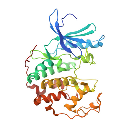
3PXY

CDK2 in complex with inhibitor JWS648


Experimental Data Snapshot

  • Method: X-RAY DIFFRACTION
  • Resolution: 1.80 Å
  • R-Value Free: 0.242 
  • R-Value Work: 0.211 
  • R-Value Observed: 0.211 

Starting Model: experimental
View more details

wwPDB Validation   3D Report Full Report


Ligand Structure Quality Assessment 


This is version 1.2 of the entry. See complete history


Literature

Discovery of a Potential Allosteric Ligand Binding Site in CDK2.

Betzi, S.Alam, R.Martin, M.Lubbers, D.J.Han, H.Jakkaraj, S.R.Georg, G.I.Schonbrunn, E.

(2011) ACS Chem Biol 6: 492-501

  • DOI: https://doi.org/10.1021/cb100410m
  • Primary Citation of Related Structures:  
    3PXF, 3PXQ, 3PXR, 3PXY, 3PXZ, 3PY0, 3PY1

  • PubMed Abstract: 

    Cyclin-dependent kinases (CDKs) are key regulatory enzymes in cell cycle progression and transcription. Aberrant activity of CDKs has been implicated in a number of medical conditions, and numerous small molecule CDK inhibitors have been reported as potential drug leads. However, these inhibitors exclusively bind to the ATP site, which is largely conserved among protein kinases, and clinical trials have not resulted in viable drug candidates, attributed in part to the lack of target selectivity. CDKs are unique among protein kinases, as their functionality strictly depends on association with their partner proteins, the cyclins. In an effort to identify potential target sites for disruption of the CDK-cyclin interaction, we probed the extrinsic fluorophore 8-anilino-1-naphthalene sulfonate (ANS) with human CDK2 and cyclin A using fluorescence spectroscopy and protein crystallography. ANS interacts with free CDK2 in a saturation-dependent manner with an apparent K(d) of 37 μM, and cyclin A displaced ANS from CDK2 with an EC(50) value of 0.6 μM. Co-crystal structures with ANS alone and in ternary complex with ATP site-directed inhibitors revealed two ANS molecules bound adjacent to one another, away from the ATP site, in a large pocket that extends from the DFG region above the C-helix. Binding of ANS is accompanied by substantial structural changes in CDK2, resulting in a C-helix conformation that is incompatible for cyclin A association. These findings indicate the potential of the ANS binding pocket as a new target site for allosteric inhibitors disrupting the interaction of CDKs and cyclins.


  • Organizational Affiliation

    Drug Discovery Department, H. Lee Moffitt Cancer Center and Research Institute, Tampa, Florida 33612, USA.


Macromolecules
Find similar proteins by:  (by identity cutoff)  |  3D Structure
Entity ID: 1
MoleculeChains Sequence LengthOrganismDetailsImage
Cell division protein kinase 2306Homo sapiensMutation(s): 0 
Gene Names: CDK2
EC: 2.7.11.22
UniProt & NIH Common Fund Data Resources
Find proteins for P24941 (Homo sapiens)
Explore P24941 
Go to UniProtKB:  P24941
PHAROS:  P24941
GTEx:  ENSG00000123374 
Entity Groups  
Sequence Clusters30% Identity50% Identity70% Identity90% Identity95% Identity100% Identity
UniProt GroupP24941
Sequence Annotations
Expand
  • Reference Sequence
Small Molecules
Ligands 2 Unique
IDChains Name / Formula / InChI Key2D Diagram3D Interactions
JWS
Query on JWS

Download Ideal Coordinates CCD File 
B [auth A]2-(4,6-diamino-1,3,5-triazin-2-yl)-4-methoxyphenol
C10 H11 N5 O2
OQGZVJIFCDXDFS-UHFFFAOYSA-N
PO4
Query on PO4

Download Ideal Coordinates CCD File 
C [auth A]PHOSPHATE ION
O4 P
NBIIXXVUZAFLBC-UHFFFAOYSA-K
Binding Affinity Annotations 
IDSourceBinding Affinity
JWS PDBBind:  3PXY IC50: 5900 (nM) from 1 assay(s)
Experimental Data & Validation

Experimental Data

  • Method: X-RAY DIFFRACTION
  • Resolution: 1.80 Å
  • R-Value Free: 0.242 
  • R-Value Work: 0.211 
  • R-Value Observed: 0.211 
  • Space Group: P 21 21 21
Unit Cell:
Length ( Å )Angle ( ˚ )
a = 52.82α = 90
b = 71.74β = 90
c = 72.07γ = 90
Software Package:
Software NamePurpose
StructureStudiodata collection
CNSrefinement
XDSdata reduction
XDSdata scaling
CNSphasing

Structure Validation

View Full Validation Report


Ligand Structure Quality Assessment 


Entry History 

Deposition Data

Revision History  (Full details and data files)

  • Version 1.0: 2011-02-16
    Type: Initial release
  • Version 1.1: 2011-07-13
    Changes: Version format compliance
  • Version 1.2: 2023-09-13
    Changes: Data collection, Database references, Derived calculations, Refinement description