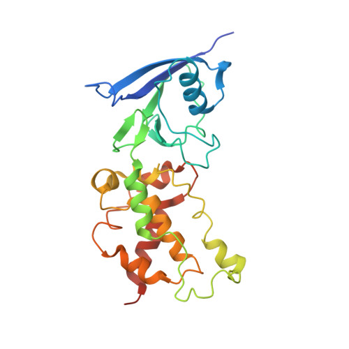
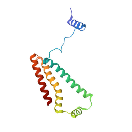
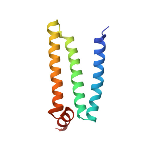
3SFE

crystal structure of porcine mitochondrial respiratory complex II bound with oxaloacetate and thiabendazole


Experimental Data Snapshot

  • Method: X-RAY DIFFRACTION
  • Resolution: 2.81 Å
  • R-Value Free: 0.270 
  • R-Value Work: 0.214 
  • R-Value Observed: 0.217 

Starting Model: experimental
View more details

wwPDB Validation   3D Report Full Report


Ligand Structure Quality Assessment 


This is version 1.2 of the entry. See complete history


Literature

Thiabendazole inhibits ubiquinone reduction activity of mitochondrial respiratory complex II via a water molecule mediated binding feature.

Zhou, Q.Zhai, Y.Lou, J.Liu, M.Pang, X.Sun, F.

(2011) Protein Cell 2: 531-542

  • DOI: https://doi.org/10.1007/s13238-011-1079-1
  • Primary Citation of Related Structures:  
    3SFD, 3SFE

  • PubMed Abstract: 

    The mitochondrial respiratory complex II or succinate: ubiquinone oxidoreductase (SQR) is a key membrane complex in both the tricarboxylic acid cycle and aerobic respiration. Five disinfectant compounds were investigated with their potent inhibition effects on the ubiquinone reduction activity of the porcine mitochondrial SQR by enzymatic assay and crystallography. Crystal structure of the SQR bound with thiabendazole (TBZ) reveals a different inhibitor-binding feature at the ubiquinone binding site where a water molecule plays an important role. The obvious inhibitory effect of TBZ based on the biochemical data (IC(50) ~100 μmol/L) and the significant structure-based binding affinity calculation (~94 μmol/L) draw the suspicion of using TBZ as a good disinfectant compound for nematode infections treatment and fruit storage.


  • Organizational Affiliation

    National Laboratory of Biomacromolecules, Institute of Biophysics, Chinese Academy of Sciences, Beijing 100101, China.


Macromolecules
Find similar proteins by:  (by identity cutoff)  |  3D Structure
Entity ID: 1
MoleculeChains Sequence LengthOrganismDetailsImage
Succinate dehydrogenase [ubiquinone] flavoprotein subunit, mitochondrial622Sus scrofaMutation(s): 0 
EC: 1.3.5.1 (PDB Primary Data), 1.1.5 (UniProt)
Membrane Entity: Yes 
UniProt
Find proteins for Q0QF01 (Sus scrofa)
Explore Q0QF01 
Go to UniProtKB:  Q0QF01
Entity Groups  
Sequence Clusters30% Identity50% Identity70% Identity90% Identity95% Identity100% Identity
UniProt GroupQ0QF01
Sequence Annotations
Expand
  • Reference Sequence
Find similar proteins by:  (by identity cutoff)  |  3D Structure
Entity ID: 2
MoleculeChains Sequence LengthOrganismDetailsImage
Succinate dehydrogenase [ubiquinone] iron-sulfur subunit, mitochondrial252Sus scrofaMutation(s): 0 
EC: 1.3.5.1 (PDB Primary Data), 1.1.5 (UniProt)
Membrane Entity: Yes 
UniProt
Find proteins for Q007T0 (Sus scrofa)
Explore Q007T0 
Go to UniProtKB:  Q007T0
Entity Groups  
Sequence Clusters30% Identity50% Identity70% Identity90% Identity95% Identity100% Identity
UniProt GroupQ007T0
Sequence Annotations
Expand
  • Reference Sequence
Find similar proteins by:  (by identity cutoff)  |  3D Structure
Entity ID: 3
MoleculeChains Sequence LengthOrganismDetailsImage
Succinate dehydrogenase cytochrome b560 subunit, mitochondrial140Sus scrofaMutation(s): 0 
Membrane Entity: Yes 
UniProt
Find proteins for D0VWV4 (Sus scrofa)
Explore D0VWV4 
Go to UniProtKB:  D0VWV4
Entity Groups  
Sequence Clusters30% Identity50% Identity70% Identity90% Identity95% Identity100% Identity
UniProt GroupD0VWV4
Sequence Annotations
Expand
  • Reference Sequence
Find similar proteins by:  (by identity cutoff)  |  3D Structure
Entity ID: 4
MoleculeChains Sequence LengthOrganismDetailsImage
Succinate dehydrogenase [ubiquinone] cytochrome b small subunit, mitochondrial103Sus scrofaMutation(s): 0 
Membrane Entity: Yes 
UniProt
Find proteins for A5GZW8 (Sus scrofa)
Explore A5GZW8 
Go to UniProtKB:  A5GZW8
Entity Groups  
Sequence Clusters30% Identity50% Identity70% Identity90% Identity95% Identity100% Identity
UniProt GroupA5GZW8
Sequence Annotations
Expand
  • Reference Sequence
Small Molecules
Ligands 7 Unique
IDChains Name / Formula / InChI Key2D Diagram3D Interactions
FAD
Query on FAD

Download Ideal Coordinates CCD File 
E [auth A]FLAVIN-ADENINE DINUCLEOTIDE
C27 H33 N9 O15 P2
VWWQXMAJTJZDQX-UYBVJOGSSA-N
HEM
Query on HEM

Download Ideal Coordinates CCD File 
J [auth C]PROTOPORPHYRIN IX CONTAINING FE
C34 H32 Fe N4 O4
KABFMIBPWCXCRK-RGGAHWMASA-L
SF4
Query on SF4

Download Ideal Coordinates CCD File 
H [auth B]IRON/SULFUR CLUSTER
Fe4 S4
LJBDFODJNLIPKO-UHFFFAOYSA-N
F3S
Query on F3S

Download Ideal Coordinates CCD File 
I [auth B]FE3-S4 CLUSTER
Fe3 S4
FCXHZBQOKRZXKS-UHFFFAOYSA-N
TMG
Query on TMG

Download Ideal Coordinates CCD File 
K [auth C]2-(1,3-THIAZOL-4-YL)-1H-BENZIMIDAZOLE
C10 H7 N3 S
WJCNZQLZVWNLKY-UHFFFAOYSA-N
FES
Query on FES

Download Ideal Coordinates CCD File 
G [auth B]FE2/S2 (INORGANIC) CLUSTER
Fe2 S2
NIXDOXVAJZFRNF-UHFFFAOYSA-N
OAA
Query on OAA

Download Ideal Coordinates CCD File 
F [auth A]OXALOACETATE ION
C4 H3 O5
KHPXUQMNIQBQEV-UHFFFAOYSA-M
Experimental Data & Validation

Experimental Data

  • Method: X-RAY DIFFRACTION
  • Resolution: 2.81 Å
  • R-Value Free: 0.270 
  • R-Value Work: 0.214 
  • R-Value Observed: 0.217 
  • Space Group: P 21 21 21
Unit Cell:
Length ( Å )Angle ( ˚ )
a = 70.71α = 90
b = 83.42β = 90
c = 294.01γ = 90
Software Package:
Software NamePurpose
HKL-2000data collection
PHASESphasing
REFMACrefinement
HKL-2000data reduction
HKL-2000data scaling

Structure Validation

View Full Validation Report


Ligand Structure Quality Assessment 


Entry History 

Deposition Data

Revision History  (Full details and data files)

  • Version 1.0: 2011-09-07
    Type: Initial release
  • Version 1.1: 2023-11-01
    Changes: Data collection, Database references, Derived calculations, Refinement description
  • Version 1.2: 2024-10-30
    Changes: Structure summary