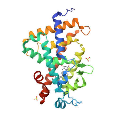
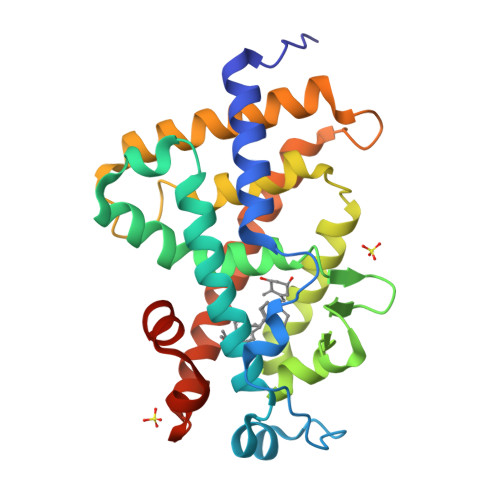
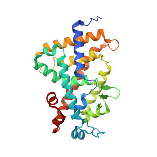
3TKC

Design, Synthesis, Evaluation and Structure of Vitamin D Analogues with Furan Side Chains


Experimental Data Snapshot

  • Method: X-RAY DIFFRACTION
  • Resolution: 1.75 Å
  • R-Value Free: 0.183 
  • R-Value Work: 0.158 
  • R-Value Observed: 0.159 

Starting Model: experimental
View more details

wwPDB Validation   3D Report Full Report


Ligand Structure Quality Assessment 


This is version 1.1 of the entry. See complete history


Literature

Design, synthesis, evaluation, and structure of vitamin D analogues with furan side chains.

Fraga, R.Zacconi, F.Sussman, F.Ordonez-Moran, P.Munoz, A.Huet, T.Molnar, F.Moras, D.Rochel, N.Maestro, M.Mourino, A.

(2012) Chemistry 18: 603-612

  • DOI: https://doi.org/10.1002/chem.201102695
  • Primary Citation of Related Structures:  
    3TKC

  • PubMed Abstract: 

    Based on the crystal structures of human vitamin D receptor (hVDR) bound to 1α,25-dihydroxy-vitamin D(3) (1,25 D) and superagonist ligands, we previously designed new superagonist ligands with a tetrahydrofuran ring at the side chain that optimize the aliphatic side-chain conformation through an entropy benefit. Following a similar strategy, four novel vitamin D analogues with aromatic furan side chains (3a, 3b, 4a, 4b) have now been developed. The triene system has been constructed by an efficient stereoselective intramolecular cyclization of an enol triflate (A-ring precursor) followed by a Suzuki-Miyaura coupling of the resulting intermediate with an alkenyl boronic ester (CD-side chain, upper fragment). The furan side chains have been constructed by gold chemistry. These analogues exhibit significant pro-differentiation effects and transactivation potency. The crystal structure of 3a in a complex with the ligand-binding domain of hVDR revealed that the side-chain furanic ring adopts two conformations.


  • Organizational Affiliation

    Departamento de Química Orgánica, Universidad de Santiago y Unidad Asociada al CSIC, Avda de las Ciencias s/n, 15782 Santiago de Compostela, Spain.


Macromolecules
Find similar proteins by:  (by identity cutoff)  |  3D Structure
Entity ID: 1
MoleculeChains Sequence LengthOrganismDetailsImage
Vitamin D3 receptor259Homo sapiensMutation(s): 0 
Gene Names: VDRNR1I1
UniProt & NIH Common Fund Data Resources
Find proteins for P11473 (Homo sapiens)
Explore P11473 
Go to UniProtKB:  P11473
PHAROS:  P11473
GTEx:  ENSG00000111424 
Entity Groups  
Sequence Clusters30% Identity50% Identity70% Identity90% Identity95% Identity100% Identity
UniProt GroupP11473
Sequence Annotations
Expand
  • Reference Sequence
Experimental Data & Validation

Experimental Data

  • Method: X-RAY DIFFRACTION
  • Resolution: 1.75 Å
  • R-Value Free: 0.183 
  • R-Value Work: 0.158 
  • R-Value Observed: 0.159 
  • Space Group: P 21 21 21
Unit Cell:
Length ( Å )Angle ( ˚ )
a = 45.018α = 90
b = 51.622β = 90
c = 132.818γ = 90
Software Package:
Software NamePurpose
MxCuBEdata collection
PHENIXmodel building
PHENIXrefinement
HKL-2000data reduction
HKL-2000data scaling
PHENIXphasing

Structure Validation

View Full Validation Report


Ligand Structure Quality Assessment 


Entry History 

Deposition Data

Revision History  (Full details and data files)

  • Version 1.0: 2012-03-07
    Type: Initial release
  • Version 1.1: 2023-09-13
    Changes: Data collection, Database references, Derived calculations, Refinement description