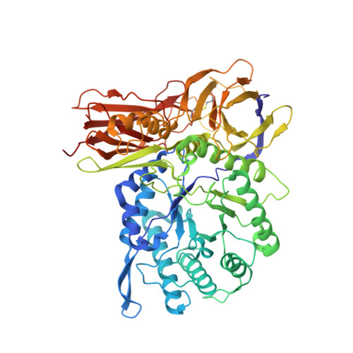
3W81

Human alpha-l-iduronidase


Experimental Data Snapshot

  • Method: X-RAY DIFFRACTION
  • Resolution: 2.30 Å
  • R-Value Free: 0.210 
  • R-Value Work: 0.169 
  • R-Value Observed: 0.171 

wwPDB Validation   3D Report Full Report


Ligand Structure Quality Assessment 


This is version 2.1 of the entry. See complete history


Literature

Human alpha-L-iduronidase uses its own N-glycan as a substrate-binding and catalytic module

Maita, N.Tsukimura, T.Taniguchi, T.Saito, S.Ohno, K.Taniguchi, H.Sakuraba, H.

(2013) Proc Natl Acad Sci U S A 110: 14628-14633

  • DOI: https://doi.org/10.1073/pnas.1306939110
  • Primary Citation of Related Structures:  
    3W81, 3W82

  • PubMed Abstract: 

    N-glycosylation is a major posttranslational modification that endows proteins with various functions. It is established that N-glycans are essential for the correct folding and stability of some enzymes; however, the actual effects of N-glycans on their activities are poorly understood. Here, we show that human α-l-iduronidase (hIDUA), of which a dysfunction causes accumulation of dermatan/heparan sulfate leading to mucopolysaccharidosis type I, uses its own N-glycan as a substrate binding and catalytic module. Structural analysis revealed that the mannose residue of the N-glycan attached to N372 constituted a part of the substrate-binding pocket and interacted directly with a substrate. A deglycosylation study showed that enzyme activity was highly correlated with the N-glycan attached to N372. The kinetics of native and deglycosylated hIDUA suggested that the N-glycan is also involved in catalytic processes. Our study demonstrates a previously unrecognized function of N-glycans.


  • Organizational Affiliation

    Laboratory of X-Ray Crystallography, Institute for Enzyme Research, University of Tokushima, Tokushima 770-8503, Japan. nmaita@tokushima-u.ac.jp


Macromolecules
Find similar proteins by:  (by identity cutoff)  |  3D Structure
Entity ID: 1
MoleculeChains Sequence LengthOrganismDetailsImage
Alpha-L-iduronidase
A, B
627Homo sapiensMutation(s): 0 
Gene Names: IDUA
EC: 3.2.1.76
UniProt & NIH Common Fund Data Resources
Find proteins for P35475 (Homo sapiens)
Explore P35475 
Go to UniProtKB:  P35475
PHAROS:  P35475
GTEx:  ENSG00000127415 
Entity Groups  
Sequence Clusters30% Identity50% Identity70% Identity90% Identity95% Identity100% Identity
UniProt GroupP35475
Glycosylation
Glycosylation Sites: 3Go to GlyGen: P35475-1
Sequence Annotations
Expand
  • Reference Sequence
Oligosaccharides

Help

Entity ID: 2
MoleculeChains Length2D Diagram Glycosylation3D Interactions
alpha-D-mannopyranose-(1-2)-alpha-D-mannopyranose-(1-3)-[alpha-D-mannopyranose-(1-2)-alpha-D-mannopyranose-(1-6)]alpha-D-mannopyranose-(1-6)-[alpha-D-mannopyranose-(1-2)-alpha-D-mannopyranose-(1-3)]beta-D-mannopyranose-(1-4)-2-acetamido-2-deoxy-beta-D-glucopyranose-(1-4)-2-acetamido-2-deoxy-beta-D-glucopyranose
C
10N-Glycosylation
Glycosylation Resources
GlyTouCan:  G89864BN
GlyCosmos:  G89864BN
GlyGen:  G89864BN
Entity ID: 3
MoleculeChains Length2D Diagram Glycosylation3D Interactions
2-acetamido-2-deoxy-beta-D-glucopyranose-(1-4)-2-acetamido-2-deoxy-beta-D-glucopyranose
D, G
2N-Glycosylation
Glycosylation Resources
GlyTouCan:  G42666HT
GlyCosmos:  G42666HT
GlyGen:  G42666HT
Entity ID: 4
MoleculeChains Length2D Diagram Glycosylation3D Interactions
alpha-D-mannopyranose-(1-2)-alpha-D-mannopyranose-(1-6)-[alpha-D-mannopyranose-(1-3)]alpha-D-mannopyranose-(1-6)-[alpha-D-mannopyranose-(1-2)-alpha-D-mannopyranose-(1-3)]beta-D-mannopyranose-(1-4)-2-acetamido-2-deoxy-beta-D-glucopyranose-(1-4)-2-acetamido-2-deoxy-beta-D-glucopyranose
E
9N-Glycosylation
Glycosylation Resources
GlyTouCan:  G83161QT
GlyCosmos:  G83161QT
GlyGen:  G83161QT
Entity ID: 5
MoleculeChains Length2D Diagram Glycosylation3D Interactions
beta-D-galactopyranose-(1-4)-2-acetamido-2-deoxy-beta-D-glucopyranose-(1-2)-alpha-D-mannopyranose-(1-6)-beta-D-mannopyranose-(1-4)-2-acetamido-2-deoxy-beta-D-glucopyranose-(1-4)-[alpha-L-fucopyranose-(1-6)]2-acetamido-2-deoxy-beta-D-glucopyranose
F
7N-Glycosylation
Glycosylation Resources
GlyTouCan:  G77653XA
GlyCosmos:  G77653XA
GlyGen:  G77653XA
Small Molecules
Ligands 6 Unique
IDChains Name / Formula / InChI Key2D Diagram3D Interactions
NAG
Query on NAG

Download Ideal Coordinates CCD File 
H [auth A],
O [auth B],
P [auth B]
2-acetamido-2-deoxy-beta-D-glucopyranose
C8 H15 N O6
OVRNDRQMDRJTHS-FMDGEEDCSA-N
TLA
Query on TLA

Download Ideal Coordinates CCD File 
I [auth A],
J [auth A],
K [auth A],
Q [auth B]
L(+)-TARTARIC ACID
C4 H6 O6
FEWJPZIEWOKRBE-JCYAYHJZSA-N
PO4
Query on PO4

Download Ideal Coordinates CCD File 
M [auth A],
T [auth B],
U [auth B],
V [auth B]
PHOSPHATE ION
O4 P
NBIIXXVUZAFLBC-UHFFFAOYSA-K
GOL
Query on GOL

Download Ideal Coordinates CCD File 
L [auth A],
R [auth B]
GLYCEROL
C3 H8 O3
PEDCQBHIVMGVHV-UHFFFAOYSA-N
PO3
Query on PO3

Download Ideal Coordinates CCD File 
S [auth B]PHOSPHITE ION
O3 P
AQSJGOWTSHOLKH-UHFFFAOYSA-N
CL
Query on CL

Download Ideal Coordinates CCD File 
N [auth A],
W [auth B]
CHLORIDE ION
Cl
VEXZGXHMUGYJMC-UHFFFAOYSA-M
Experimental Data & Validation

Experimental Data

  • Method: X-RAY DIFFRACTION
  • Resolution: 2.30 Å
  • R-Value Free: 0.210 
  • R-Value Work: 0.169 
  • R-Value Observed: 0.171 
  • Space Group: H 3
Unit Cell:
Length ( Å )Angle ( ˚ )
a = 259.225α = 90
b = 259.225β = 90
c = 71.835γ = 120
Software Package:
Software NamePurpose
ADSCdata collection
SHARPphasing
REFMACrefinement
HKL-2000data reduction
HKL-2000data scaling

Structure Validation

View Full Validation Report


Ligand Structure Quality Assessment 


Entry History 

Deposition Data

Revision History  (Full details and data files)

  • Version 1.0: 2013-08-21
    Type: Initial release
  • Version 1.1: 2013-10-16
    Changes: Database references
  • Version 2.0: 2020-07-29
    Type: Remediation
    Reason: Carbohydrate remediation
    Changes: Atomic model, Data collection, Derived calculations, Structure summary
  • Version 2.1: 2024-11-20
    Changes: Data collection, Database references, Structure summary