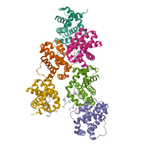
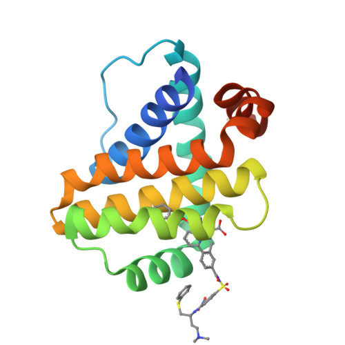
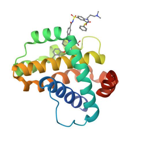
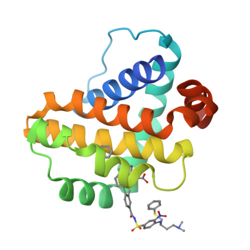
3WIY

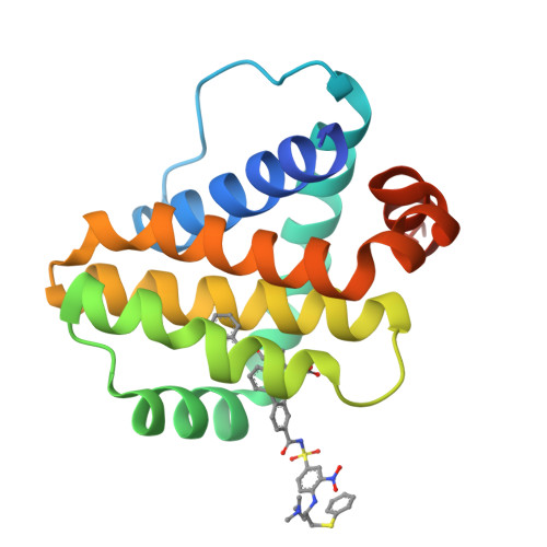
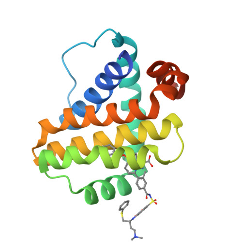
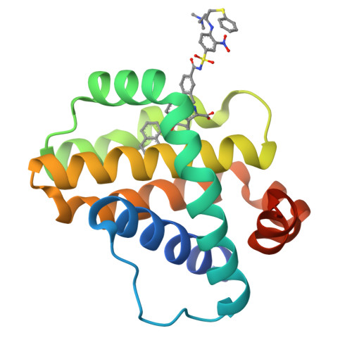
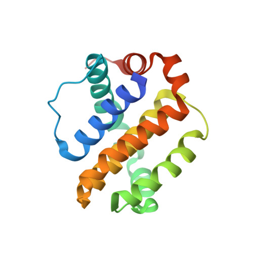
Crystal structure of Mcl-1 in complex with compound 10


Experimental Data Snapshot

  • Method: X-RAY DIFFRACTION
  • Resolution: 2.15 Å
  • R-Value Free: 0.283 
  • R-Value Work: 0.211 
  • R-Value Observed: 0.214 

Starting Model: experimental
View more details

wwPDB Validation   3D Report Full Report


Ligand Structure Quality Assessment 


This is version 1.2 of the entry. See complete history


Literature

Discovery of potent Mcl-1/Bcl-xL dual inhibitors by using a hybridization strategy based on structural analysis of target proteins.

Tanaka, Y.Aikawa, K.Nishida, G.Homma, M.Sogabe, S.Igaki, S.Hayano, Y.Sameshima, T.Miyahisa, I.Kawamoto, T.Tawada, M.Imai, Y.Inazuka, M.Cho, N.Imaeda, Y.Ishikawa, T.

(2013) J Med Chem 56: 9635-9645

  • DOI: https://doi.org/10.1021/jm401170c
  • Primary Citation of Related Structures:  
    3WIX, 3WIY, 3WIZ

  • PubMed Abstract: 

    Mcl-1 and Bcl-xL are crucial regulators of apoptosis, therefore dual inhibitors of both proteins could serve as promising new anticancer drugs. To design Mcl-1/Bcl-xL dual inhibitors, we performed structure-guided analyses of the corresponding selective Mcl-1 and Bcl-xL inhibitors. A cocrystal structure of a pyrazolo[1,5-a]pyridine derivative with Mcl-1 protein was successfully determined and revealed the protein-ligand binding mode. The key structure for Bcl-xL inhibition was further confirmed through the substructural analysis of ABT-263, a representative Bcl-xL/Bcl-2/Bcl-w inhibitor developed by Abbott Laboratories. On the basis of the structural data from this analysis, we designed hybrid compounds by tethering the Mcl-1 and Bcl-xL inhibitors together. The results of X-ray crystallographic analysis of hybrid compound 10 in complexes with both Mcl-1 and Bcl-xL demonstrated its binding mode with each protein. Following further optimization, compound 11 showed potent Mcl-1/Bcl-xL dual inhibitory activity (Mcl-1, IC50 = 0.088 μM; and Bcl-xL, IC50 = 0.0037 μM).


  • Organizational Affiliation

    Pharmaceutical Research Division, Takeda Pharmaceutical Company Limited , 26-1, Muraoka-Higashi 2-chome, Fujisawa, Kanagawa 251-8555, Japan.


Macromolecules
Find similar proteins by:  (by identity cutoff)  |  3D Structure
Entity ID: 1
MoleculeChains Sequence LengthOrganismDetailsImage
Induced myeloid leukemia cell differentiation protein Mcl-1
A, B, C, D, E
A, B, C, D, E, F
161Homo sapiensMutation(s): 0 
Gene Names: MCL1BCL2L3
UniProt & NIH Common Fund Data Resources
Find proteins for Q07820 (Homo sapiens)
Explore Q07820 
Go to UniProtKB:  Q07820
PHAROS:  Q07820
GTEx:  ENSG00000143384 
Entity Groups  
Sequence Clusters30% Identity50% Identity70% Identity90% Identity95% Identity100% Identity
UniProt GroupQ07820
Sequence Annotations
Expand
  • Reference Sequence
Small Molecules
Binding Affinity Annotations 
IDSourceBinding Affinity
LC6 PDBBind:  3WIY IC50: 610 (nM) from 1 assay(s)
BindingDB:  3WIY IC50: 610 (nM) from 1 assay(s)
Experimental Data & Validation

Experimental Data

  • Method: X-RAY DIFFRACTION
  • Resolution: 2.15 Å
  • R-Value Free: 0.283 
  • R-Value Work: 0.211 
  • R-Value Observed: 0.214 
  • Space Group: P 1
Unit Cell:
Length ( Å )Angle ( ˚ )
a = 37.288α = 101.92
b = 65.498β = 89.83
c = 97.773γ = 99.26
Software Package:
Software NamePurpose
MOLREPphasing
REFMACrefinement
HKL-2000data reduction
HKL-2000data scaling

Structure Validation

View Full Validation Report


Ligand Structure Quality Assessment 


Entry History 

Deposition Data

Revision History  (Full details and data files)

  • Version 1.0: 2013-11-27
    Type: Initial release
  • Version 1.1: 2014-03-12
    Changes: Database references
  • Version 1.2: 2023-11-08
    Changes: Data collection, Database references, Derived calculations, Refinement description