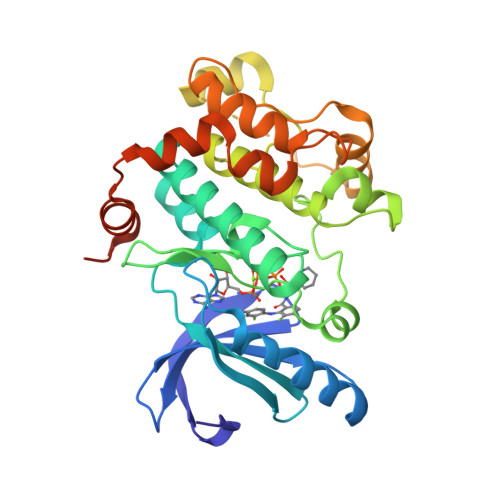
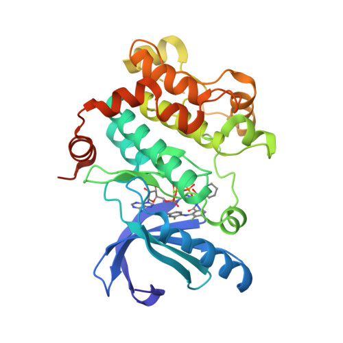
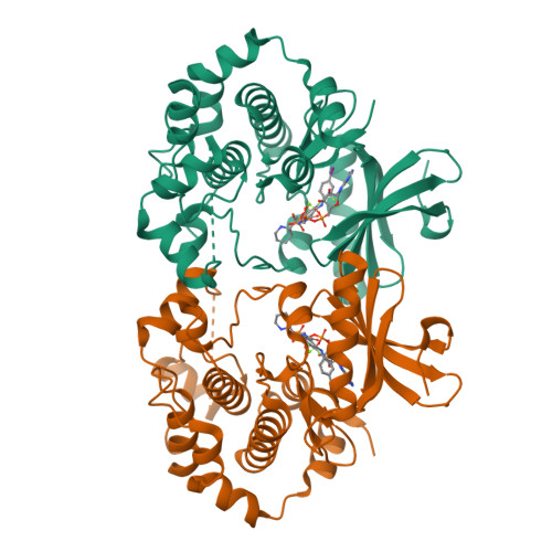
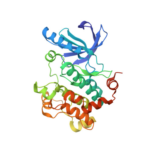
4LMN

Crystal Structure of MEK1 kinase bound to GDC0973


Experimental Data Snapshot

  • Method: X-RAY DIFFRACTION
  • Resolution: 2.80 Å
  • R-Value Free: 0.233 
  • R-Value Work: 0.191 
  • R-Value Observed: 0.193 

wwPDB Validation   3D Report Full Report


Ligand Structure Quality Assessment 


This is version 1.3 of the entry. See complete history


Literature

Mechanism of MEK inhibition determines efficacy in mutant KRAS- versus BRAF-driven cancers.

Hatzivassiliou, G.Haling, J.R.Chen, H.Song, K.Price, S.Heald, R.Hewitt, J.F.Zak, M.Peck, A.Orr, C.Merchant, M.Hoeflich, K.P.Chan, J.Luoh, S.M.Anderson, D.J.Ludlam, M.J.Wiesmann, C.Ultsch, M.Friedman, L.S.Malek, S.Belvin, M.

(2013) Nature 501: 232-236

  • DOI: https://doi.org/10.1038/nature12441
  • Primary Citation of Related Structures:  
    4LMN

  • PubMed Abstract: 

    KRAS and BRAF activating mutations drive tumorigenesis through constitutive activation of the MAPK pathway. As these tumours represent an area of high unmet medical need, multiple allosteric MEK inhibitors, which inhibit MAPK signalling in both genotypes, are being tested in clinical trials. Impressive single-agent activity in BRAF-mutant melanoma has been observed; however, efficacy has been far less robust in KRAS-mutant disease. Here we show that, owing to distinct mechanisms regulating MEK activation in KRAS- versus BRAF-driven tumours, different mechanisms of inhibition are required for optimal antitumour activity in each genotype. Structural and functional analysis illustrates that MEK inhibitors with superior efficacy in KRAS-driven tumours (GDC-0623 and G-573, the former currently in phase I clinical trials) form a strong hydrogen-bond interaction with S212 in MEK that is critical for blocking MEK feedback phosphorylation by wild-type RAF. Conversely, potent inhibition of active, phosphorylated MEK is required for strong inhibition of the MAPK pathway in BRAF-mutant tumours, resulting in superior efficacy in this genotype with GDC-0973 (also known as cobimetinib), a MEK inhibitor currently in phase III clinical trials. Our study highlights that differences in the activation state of MEK in KRAS-mutant tumours versus BRAF-mutant tumours can be exploited through the design of inhibitors that uniquely target these distinct activation states of MEK. These inhibitors are currently being evaluated in clinical trials to determine whether improvements in therapeutic index within KRAS versus BRAF preclinical models translate to improved clinical responses in patients.


  • Organizational Affiliation

    Department of Translational Oncology, Genentech, Inc., 1 DNA Way, South San Francisco, California 94080, USA. hatzivassiliou.georgia@gene.com


Macromolecules
Find similar proteins by:  (by identity cutoff)  |  3D Structure
Entity ID: 1
MoleculeChains Sequence LengthOrganismDetailsImage
Dual specificity mitogen-activated protein kinase kinase 1341Homo sapiensMutation(s): 0 
Gene Names: MAP2K1MEK1PRKMK1
EC: 2.7.12.2
UniProt & NIH Common Fund Data Resources
Find proteins for Q02750 (Homo sapiens)
Explore Q02750 
Go to UniProtKB:  Q02750
PHAROS:  Q02750
GTEx:  ENSG00000169032 
Entity Groups  
Sequence Clusters30% Identity50% Identity70% Identity90% Identity95% Identity100% Identity
UniProt GroupQ02750
Sequence Annotations
Expand
  • Reference Sequence
Small Molecules
Ligands 3 Unique
IDChains Name / Formula / InChI Key2D Diagram3D Interactions
EUI
Query on EUI

Download Ideal Coordinates CCD File 
D [auth A][3,4-BIS(FLUORANYL)-2-[(2-FLUORANYL-4-IODANYL-PHENYL)AMINO]PHENYL]-[3-OXIDANYL-3-[(2S)-PIPERIDIN-2-YL]AZETIDIN-1-YL]METHANONE
C21 H21 F3 I N3 O2
BSMCAPRUBJMWDF-KRWDZBQOSA-N
ANP
Query on ANP

Download Ideal Coordinates CCD File 
B [auth A]PHOSPHOAMINOPHOSPHONIC ACID-ADENYLATE ESTER
C10 H17 N6 O12 P3
PVKSNHVPLWYQGJ-KQYNXXCUSA-N
MG
Query on MG

Download Ideal Coordinates CCD File 
C [auth A]MAGNESIUM ION
Mg
JLVVSXFLKOJNIY-UHFFFAOYSA-N
Binding Affinity Annotations 
IDSourceBinding Affinity
EUI PDBBind:  4LMN Ki: 0.05 (nM) from 1 assay(s)
Experimental Data & Validation

Experimental Data

  • Method: X-RAY DIFFRACTION
  • Resolution: 2.80 Å
  • R-Value Free: 0.233 
  • R-Value Work: 0.191 
  • R-Value Observed: 0.193 
  • Space Group: P 62
Unit Cell:
Length ( Å )Angle ( ˚ )
a = 81.61α = 90
b = 81.61β = 90
c = 129.809γ = 120
Software Package:
Software NamePurpose
HKL-2000data collection
AMoREphasing
REFMACrefinement
HKL-2000data reduction
SCALEPACKdata scaling
BUSTERrefinement

Structure Validation

View Full Validation Report


Ligand Structure Quality Assessment 


Entry History 

Deposition Data

  • Released Date: 2013-08-07 
  • Deposition Author(s): Ultsch, M.H.

Revision History  (Full details and data files)

  • Version 1.0: 2013-08-07
    Type: Initial release
  • Version 1.1: 2013-08-28
    Changes: Database references
  • Version 1.2: 2013-09-25
    Changes: Database references
  • Version 1.3: 2024-02-28
    Changes: Data collection, Database references, Derived calculations