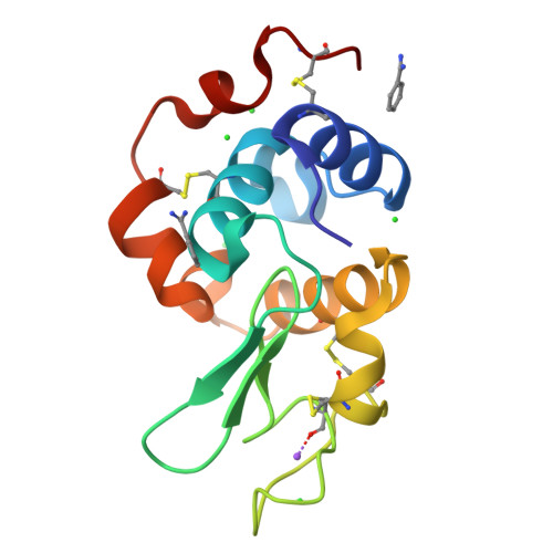
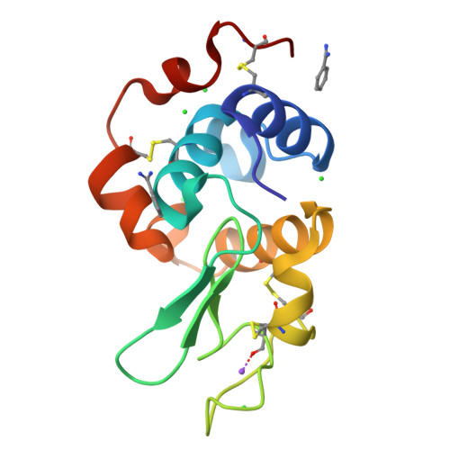
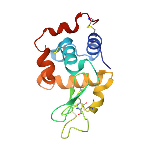
Hitting the target: fragment screening with acoustic in situ co-crystallization of proteins plus fragment libraries on pin-mounted data-collection micromeshes.
Yin, X., Scalia, A., Leroy, L., Cuttitta, C.M., Polizzo, G.M., Ericson, D.L., Roessler, C.G., Campos, O., Ma, M.Y., Agarwal, R., Jackimowicz, R., Allaire, M., Orville, A.M., Sweet, R.M., Soares, A.S.(2014) Acta Crystallogr D Biol Crystallogr 70: 1177-1189
- PubMed: 24816088
- DOI: https://doi.org/10.1107/S1399004713034603
- Primary Citation of Related Structures:
4N8Z, 4NCY - PubMed Abstract:
Acoustic droplet ejection (ADE) is a powerful technology that supports crystallographic applications such as growing, improving and manipulating protein crystals. A fragment-screening strategy is described that uses ADE to co-crystallize proteins with fragment libraries directly on MiTeGen MicroMeshes. Co-crystallization trials can be prepared rapidly and economically. The high speed of specimen preparation and the low consumption of fragment and protein allow the use of individual rather than pooled fragments. The Echo 550 liquid-handling instrument (Labcyte Inc., Sunnyvale, California, USA) generates droplets with accurate trajectories, which allows multiple co-crystallization experiments to be discretely positioned on a single data-collection micromesh. This accuracy also allows all components to be transferred through small apertures. Consequently, the crystallization tray is in equilibrium with the reservoir before, during and after the transfer of protein, precipitant and fragment to the micromesh on which crystallization will occur. This strict control of the specimen environment means that the crystallography experiments remain identical as the working volumes are decreased from the few microlitres level to the few nanolitres level. Using this system, lysozyme, thermolysin, trypsin and stachydrine demethylase crystals were co-crystallized with a small 33-compound mini-library to search for fragment hits. This technology pushes towards a much faster, more automated and more flexible strategy for structure-based drug discovery using as little as 2.5 nl of each major component.
Organizational Affiliation:
Office of Educational Programs, Brookhaven National Laboratory, Upton, NY 11973-5000, USA.


