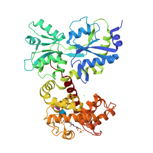
Crystal structure of MltF from Pseudomonas aeruginosa complexed with bulgecin and muropeptide
Redden, E., Thunnissen, A.M.W.H.To be published.
Experimental Data Snapshot
Entity ID: 1 | |||||
|---|---|---|---|---|---|
| Molecule | Chains | Sequence Length | Organism | Details | Image |
| Membrane-bound lytic murein transglycosylase F | 435 | Pseudomonas aeruginosa PAO1 | Mutation(s): 0 Gene Names: mltF, PA3764 EC: 4.2.2 | 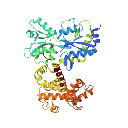 | |
UniProt | |||||
Find proteins for Q9HXN1 (Pseudomonas aeruginosa (strain ATCC 15692 / DSM 22644 / CIP 104116 / JCM 14847 / LMG 12228 / 1C / PRS 101 / PAO1)) Explore Q9HXN1 Go to UniProtKB: Q9HXN1 | |||||
Entity Groups | |||||
| Sequence Clusters | 30% Identity50% Identity70% Identity90% Identity95% Identity100% Identity | ||||
| UniProt Group | Q9HXN1 | ||||
Sequence AnnotationsExpand | |||||
| |||||
| Ligands 3 Unique | |||||
|---|---|---|---|---|---|
| ID | Chains | Name / Formula / InChI Key | 2D Diagram | 3D Interactions | |
| BLG Query on BLG | C [auth A] | 4-O-(4-O-SULFONYL-N-ACETYLGLUCOSAMININYL)-5-METHYLHYDROXY-L-PROLINE-TAURINE C16 H30 N3 O14 S2 RPNZWZDLNYCCIG-HMMVDTEZSA-O |  | ||
| CL Query on CL | G [auth A], H [auth A], I [auth A] | CHLORIDE ION Cl VEXZGXHMUGYJMC-UHFFFAOYSA-M |  | ||
| MG Query on MG | D [auth A], E [auth A], F [auth A] | MAGNESIUM ION Mg JLVVSXFLKOJNIY-UHFFFAOYSA-N |  | ||
| Length ( Å ) | Angle ( ˚ ) |
|---|---|
| a = 58.325 | α = 90 |
| b = 82.239 | β = 90 |
| c = 95.207 | γ = 90 |
| Software Name | Purpose |
|---|---|
| MOSFLM | data reduction |
| SCALA | data scaling |
| WARP | model building |
| PHENIX | refinement |
| PDB_EXTRACT | data extraction |