4YDG | pdb_00004ydg

Crystal structure of compound 10 in complex with HTLV-1 Protease


Experimental Data Snapshot

  • Method: X-RAY DIFFRACTION
  • Resolution: 3.25 Å
  • R-Value Free: 
    0.317 (Depositor), 0.320 (DCC) 
  • R-Value Work: 
    0.279 (Depositor), 0.280 (DCC) 
  • R-Value Observed: 
    0.281 (Depositor) 

Starting Model: experimental
View more details

wwPDB Validation   3D Report Full Report


Ligand Structure Quality Assessment 

Created with Raphaël 2.3.0Worse 01 BetterLigand structure goodness of fit to experimental dataBest fitted G3GClick on this verticalbar to view details

This is version 1.1 of the entry. See complete history


Literature

Privileged Structures Meet Human T-Cell Leukemia Virus-1 (HTLV-1): C2-Symmetric 3,4-Disubstituted Pyrrolidines as Nonpeptidic HTLV-1 Protease Inhibitors.

Kuhnert, M.Blum, A.Steuber, H.Diederich, W.E.

(2015) J Med Chem 58: 4845-4850

  • DOI: https://doi.org/10.1021/acs.jmedchem.5b00346
  • Primary Citation of Related Structures:  
    4YDF, 4YDG

  • PubMed Abstract: 

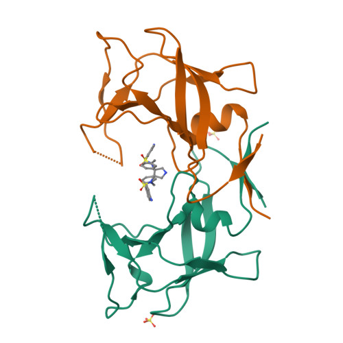
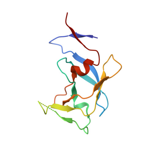
    3,4-disubstituted pyrrolidines originally designed to inhibit the closely related HIV-1 protease were evaluated as privileged structures against HTLV-1 protease (HTLV-1 PR). The most potent inhibitor of this series exhibits two-digit nanomolar affinity and represents, to the best of our knowledge, the most potent nonpeptidic inhibitor of HTLV-1 PR described so far. The X-ray structures of two representatives bound to HTLV-1 PR were determined, and the structural basis of their affinity is discussed.


  • Organizational Affiliation

    †Institut für Pharmazeutische Chemie, Philipps-Universität Marburg, Hans-Meerwein-Straße 3, 35043 Marburg, Germany.


Macromolecules
Find similar proteins by:  (by identity cutoff)  |  3D Structure
Entity ID: 1
MoleculeChains Sequence LengthOrganismDetailsImage
HTLV-1 protease
A, B
116Human T-cell lymphotrophic virus type 1 (strain ATK)Mutation(s): 1 
Gene Names: gag-pro-pol
EC: 3.4.23 (PDB Primary Data), 2.7.7.49 (PDB Primary Data), 2.7.7.7 (PDB Primary Data), 3.1.26.4 (PDB Primary Data)
UniProt
Find proteins for P03362 (Human T-cell leukemia virus 1 (strain Japan ATK-1 subtype A))
Explore P03362 
Go to UniProtKB:  P03362
Entity Groups  
Sequence Clusters30% Identity50% Identity70% Identity90% Identity95% Identity100% Identity
UniProt GroupP03362
Sequence Annotations
Expand
  • Reference Sequence
Experimental Data & Validation

Experimental Data

  • Method: X-RAY DIFFRACTION
  • Resolution: 3.25 Å
  • R-Value Free:  0.317 (Depositor), 0.320 (DCC) 
  • R-Value Work:  0.279 (Depositor), 0.280 (DCC) 
  • R-Value Observed: 0.281 (Depositor) 
Space Group: P 63 2 2
Unit Cell:
Length ( Å )Angle ( ˚ )
a = 77.61α = 90
b = 77.61β = 90
c = 160.23γ = 120
Software Package:
Software NamePurpose
PHENIXrefinement
XDSdata reduction
XSCALEdata scaling
PHASERphasing

Structure Validation

View Full Validation Report


Ligand Structure Quality Assessment 

Created with Raphaël 2.3.0Worse 01 BetterLigand structure goodness of fit to experimental dataBest fitted G3GClick on this verticalbar to view details

Entry History 

Deposition Data

Revision History  (Full details and data files)

  • Version 1.0: 2016-02-03
    Type: Initial release
  • Version 1.1: 2024-01-10
    Changes: Data collection, Database references, Refinement description