4AXX

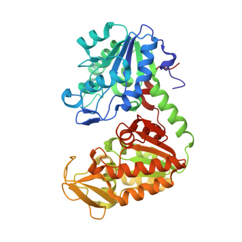
The catalytically active fully closed conformation of human phosphoglycerate kinase in complex with ADP 3-phosphoglycerate and beryllium trifluoride


Experimental Data Snapshot

  • Method: X-RAY DIFFRACTION
  • Resolution: 1.74 Å
  • R-Value Free: 0.206 
  • R-Value Work: 0.170 
  • R-Value Observed: 0.172 

Starting Model: experimental
View more details

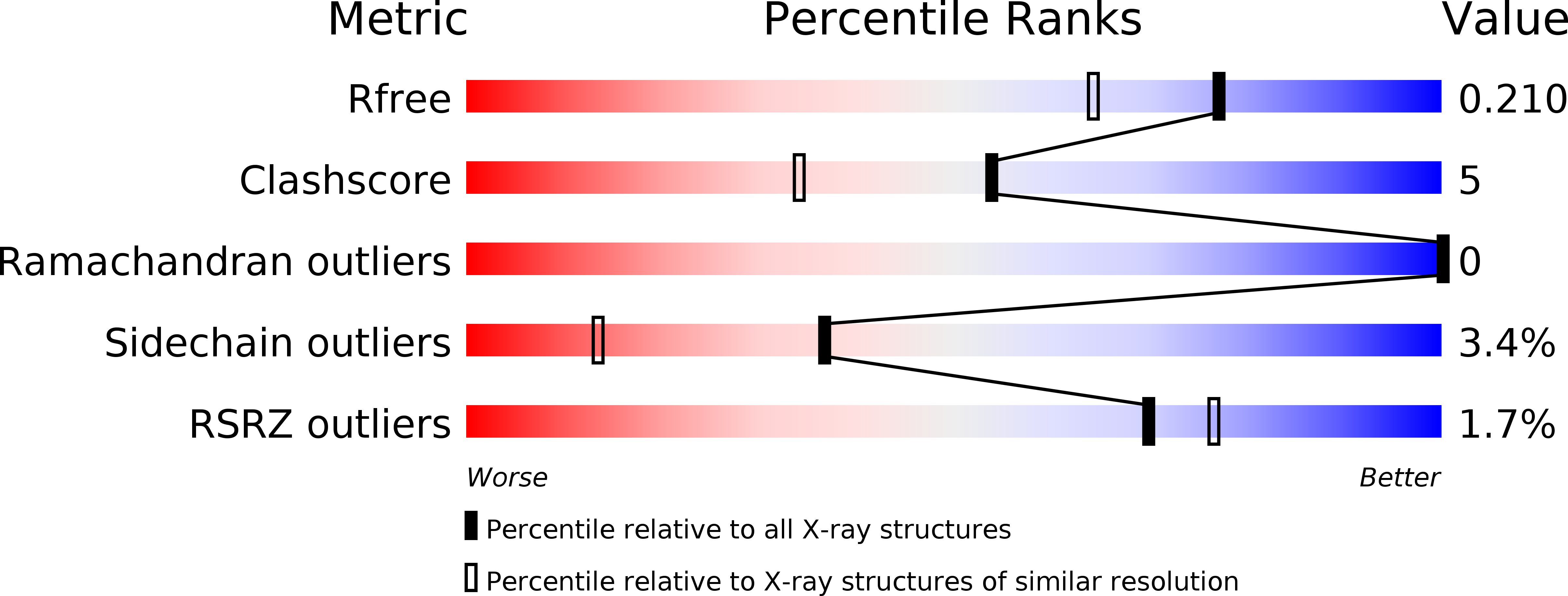
wwPDB Validation   3D Report Full Report


Ligand Structure Quality Assessment 


This is version 1.3 of the entry. See complete history


Literature

Metal fluorides-multi-functional tools for the study of phosphoryl transfer enzymes, a practical guide.

Pellegrini, E.Juyoux, P.von Velsen, J.Baxter, N.J.Dannatt, H.R.W.Jin, Y.Cliff, M.J.Waltho, J.P.Bowler, M.W.

(2024) Structure 

  • DOI: https://doi.org/10.1016/j.str.2024.07.007
  • Primary Citation of Related Structures:  
    2X13, 2X14, 3ZI4, 4AXX

  • PubMed Abstract: 

    Enzymes facilitating the transfer of phosphate groups constitute the most extensive protein families across all kingdoms of life. They make up approximately 10% of the proteins found in the human genome. Understanding the mechanisms by which enzymes catalyze these reactions is essential in characterizing the processes they regulate. Metal fluorides can be used as multifunctional tools to study these enzymes. These ionic species bear the same charge as phosphate and the transferring phosphoryl group and, in addition, allow the enzyme to be trapped in catalytically important states with spectroscopically sensitive atoms interacting directly with active site residues. The ionic nature of these phosphate surrogates also allows their removal and replacement with other analogs. Here, we describe the best practices to obtain these complexes, their use in NMR, X-ray crystallography, cryo-EM, and SAXS and describe a new metal fluoride, scandium tetrafluoride, which has significant anomalous signal using soft X-rays.


  • Organizational Affiliation

    European Molecular Biology Laboratory, 71 avenue des Martyrs, CS 90181, 38042 Grenoble, France.


Macromolecules
Find similar proteins by:  (by identity cutoff)  |  3D Structure
Entity ID: 1
MoleculeChains Sequence LengthOrganismDetailsImage
PHOSPHOGLYCERATE KINASE 1417Homo sapiensMutation(s): 0 
EC: 2.7.2.3
UniProt & NIH Common Fund Data Resources
Find proteins for P00558 (Homo sapiens)
Explore P00558 
Go to UniProtKB:  P00558
PHAROS:  P00558
GTEx:  ENSG00000102144 
Entity Groups  
Sequence Clusters30% Identity50% Identity70% Identity90% Identity95% Identity100% Identity
UniProt GroupP00558
Sequence Annotations
Expand
  • Reference Sequence
Small Molecules
Binding Affinity Annotations 
IDSourceBinding Affinity
3PG BindingDB:  4AXX Kd: 1.09e+5 (nM) from 1 assay(s)
Experimental Data & Validation

Experimental Data

  • Method: X-RAY DIFFRACTION
  • Resolution: 1.74 Å
  • R-Value Free: 0.206 
  • R-Value Work: 0.170 
  • R-Value Observed: 0.172 
  • Space Group: P 21 21 21
Unit Cell:
Length ( Å )Angle ( ˚ )
a = 38.82α = 90
b = 90.8β = 90
c = 108.54γ = 90
Software Package:
Software NamePurpose
REFMACrefinement
MOSFLMdata reduction
SCALAdata scaling
MOLREPphasing

Structure Validation

View Full Validation Report


Ligand Structure Quality Assessment 


Entry History 

Deposition Data

  • Released Date: 2013-06-26 
  • Deposition Author(s): Bowler, M.W.

Revision History  (Full details and data files)

  • Version 1.0: 2013-06-26
    Type: Initial release
  • Version 1.1: 2013-10-02
    Changes: Atomic model, Database references, Other, Structure summary
  • Version 1.2: 2023-12-20
    Changes: Data collection, Database references, Derived calculations, Other, Refinement description
  • Version 1.3: 2024-08-21
    Changes: Database references