4CD8

The structure of GH113 beta-mannanase AaManA from Alicyclobacillus acidocaldarius in complex with ManMIm


Experimental Data Snapshot

  • Method: X-RAY DIFFRACTION
  • Resolution: 1.47 Å
  • R-Value Free: 
    0.215 (Depositor), 0.230 (DCC) 
  • R-Value Work: 
    0.170 (Depositor), 0.180 (DCC) 
  • R-Value Observed: 
    0.173 (Depositor) 

Starting Model: experimental
View more details

wwPDB Validation   3D Report Full Report


Ligand Structure Quality Assessment 

Created with Raphaël 2.3.0Worse 01 BetterLigand structure goodness of fit to experimental dataBest fitted MVLClick on this verticalbar to view detailsBest fitted BMAClick on this verticalbar to view details

This is version 1.2 of the entry. See complete history


Literature

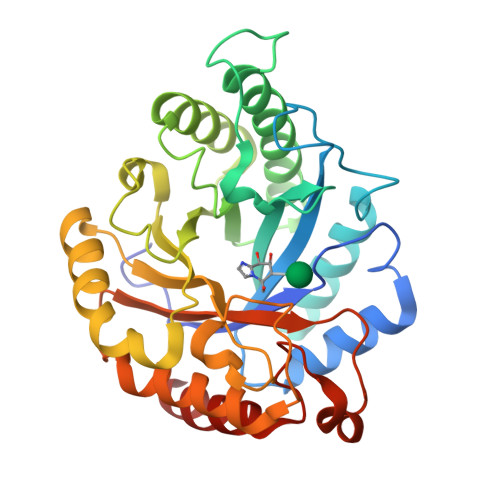
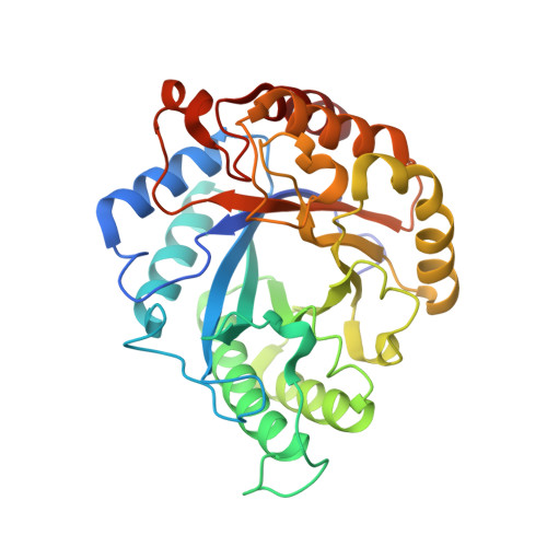
Combined Inhibitor Free-Energy Landscape and Structural Analysis Reports on the Mannosidase Conformational Coordinate.

Williams, R.J.Iglesias-Fernandez, J.Stepper, J.Jackson, A.Thompson, A.J.Lowe, E.C.White, J.M.Gilbert, H.J.Rovira, C.Davies, G.J.Williams, S.J.

(2014) Angew Chem Int Ed Engl 53: 1087

  • DOI: https://doi.org/10.1002/anie.201308334
  • Primary Citation of Related Structures:  
    4CD4, 4CD5, 4CD6, 4CD7, 4CD8

  • PubMed Abstract: 

    Mannosidases catalyze the hydrolysis of a diverse range of polysaccharides and glycoconjugates, and the various sequence-based mannosidase families have evolved ingenious strategies to overcome the stereoelectronic challenges of mannoside chemistry. Using a combination of computational chemistry, inhibitor design and synthesis, and X-ray crystallography of inhibitor/enzyme complexes, it is demonstrated that mannoimidazole-type inhibitors are energetically poised to report faithfully on mannosidase transition-state conformation, and provide direct evidence for the conformational itinerary used by diverse mannosidases, including β-mannanases from families GH26 and GH113. Isofagomine-type inhibitors are poor mimics of transition-state conformation, owing to the high energy barriers that must be crossed to attain mechanistically relevant conformations, however, these sugar-shaped heterocycles allow the acquisition of ternary complexes that span the active site, thus providing valuable insight into active-site residues involved in substrate recognition.


  • Organizational Affiliation

    School of Chemistry and Bio21 Molecular Science and Biotechnology Institute, University of Melbourne, Parkville, Vic 3010 (Australia).


Macromolecules
Find similar proteins by:  (by identity cutoff)  |  3D Structure
Entity ID: 1
MoleculeChains Sequence LengthOrganismDetailsImage
ENDO-BETA-1,4-MANNANASE320Alicyclobacillus acidocaldariusMutation(s): 0 
EC: 3.2.1.78
UniProt
Find proteins for A5H1I6 (Alicyclobacillus acidocaldarius)
Explore A5H1I6 
Go to UniProtKB:  A5H1I6
Entity Groups  
Sequence Clusters30% Identity50% Identity70% Identity90% Identity95% Identity100% Identity
UniProt GroupA5H1I6
Sequence Annotations
Expand
  • Reference Sequence
Small Molecules
Ligands 2 Unique
IDChains Name / Formula / InChI Key2D Diagram3D Interactions
MVL
Query on MVL

Download Ideal Coordinates CCD File 
B [auth A](5R,6R,7S,8R)-5-(HYDROXYMETHYL)-5,6,7,8-TETRAHYDROIMIDAZO[1,2-A]PYRIDINE-6,7,8-TRIOL
C8 H12 N2 O4
RZRDQZQPTISYKY-JWXFUTCRSA-N
BMA
Query on BMA

Download Ideal Coordinates CCD File 
C [auth A]beta-D-mannopyranose
C6 H12 O6
WQZGKKKJIJFFOK-RWOPYEJCSA-N
Experimental Data & Validation

Experimental Data

  • Method: X-RAY DIFFRACTION
  • Resolution: 1.47 Å
  • R-Value Free:  0.215 (Depositor), 0.230 (DCC) 
  • R-Value Work:  0.170 (Depositor), 0.180 (DCC) 
  • R-Value Observed: 0.173 (Depositor) 
Space Group: P 21 21 21
Unit Cell:
Length ( Å )Angle ( ˚ )
a = 43.82α = 90
b = 75.89β = 90
c = 91.48γ = 90
Software Package:
Software NamePurpose
REFMACrefinement
xia2data reduction
xia2data scaling
MOLREPphasing

Structure Validation

View Full Validation Report


Ligand Structure Quality Assessment 

Created with Raphaël 2.3.0Worse 01 BetterLigand structure goodness of fit to experimental dataBest fitted MVLClick on this verticalbar to view detailsBest fitted BMAClick on this verticalbar to view details

Entry History 

Revision History  (Full details and data files)

  • Version 1.0: 2014-04-02
    Type: Initial release
  • Version 1.1: 2020-07-29
    Type: Remediation
    Reason: Carbohydrate remediation
    Changes: Data collection, Derived calculations, Other, Structure summary
  • Version 1.2: 2023-12-20
    Changes: Data collection, Database references, Derived calculations, Refinement description, Structure summary