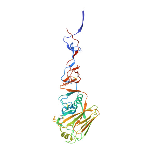
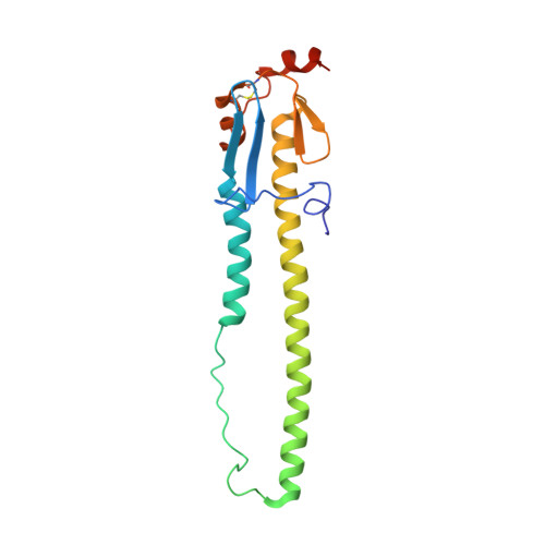
4DJ6

Structure of the hemagglutinin from a highly pathogenic H7N7 influenza virus


Experimental Data Snapshot

  • Method: X-RAY DIFFRACTION
  • Resolution: 2.61 Å
  • R-Value Free: 0.236 
  • R-Value Work: 0.203 
  • R-Value Observed: 0.205 

wwPDB Validation   3D Report Full Report


Ligand Structure Quality Assessment 


This is version 2.1 of the entry. See complete history


Literature

Structure and receptor complexes of the hemagglutinin from a highly pathogenic H7N7 influenza virus.

Yang, H.Carney, P.J.Donis, R.O.Stevens, J.

(2012) J Virol 86: 8645-8652

  • DOI: https://doi.org/10.1128/JVI.00281-12
  • Primary Citation of Related Structures:  
    4DJ6, 4DJ7, 4DJ8

  • PubMed Abstract: 

    Recurrence of highly pathogenic avian influenza (HPAI) virus subtype H7 in poultry continues to be a public health concern. In 2003, an HPAI H7N7 outbreak in The Netherlands infected 89 people in close contact with affected poultry and resulted in one fatal case. In previous studies, the virus isolated from this fatal case, A/Netherlands/219/2003 (NL219) caused a lethal infection in mouse models and had increased replication efficiency and a broader tissue distribution than nonlethal isolates from the same outbreak. A mutation which introduces a potential glycosylation site at Asn123 in the NL219 hemagglutinin was postulated to contribute to the pathogenic properties of this virus. To study this further, we have expressed the NL219 hemagglutinin in a baculovirus expression system and performed a structural analysis of the hemagglutinin in complex with avian and human receptor analogs. Glycan microarray and kinetic analysis were performed to compare the receptor binding profile of the wild-type recombinant NL219 HA to a variant with a threonine-to-alanine mutation at position 125, resulting in loss of the glycosylation site at Asn123. The results suggest that the additional glycosylation sequon increases binding affinity to avian-type α2-3-linked sialosides rather than switching to a human-like receptor specificity and highlight the mechanistic diversity of these pathogens, which calls attention to the need for further studies to fully understand the unique properties of these viruses.


  • Organizational Affiliation

    Influenza Division, National Center for Immunization and Respiratory Diseases, Centers for Disease Control and Prevention, Atlanta, Georgia, USA.


Macromolecules
Find similar proteins by:  (by identity cutoff)  |  3D Structure
Entity ID: 1
MoleculeChains Sequence LengthOrganismDetailsImage
Hemagglutinin
A, C, E
327Influenza A virus (A/Netherlands/219/2003(H7N7))Mutation(s): 0 
UniProt
Find proteins for Q6VMK1 (Influenza A virus)
Explore Q6VMK1 
Go to UniProtKB:  Q6VMK1
Entity Groups  
Sequence Clusters30% Identity50% Identity70% Identity90% Identity95% Identity100% Identity
UniProt GroupQ6VMK1
Glycosylation
Glycosylation Sites: 3
Sequence Annotations
Expand
  • Reference Sequence
Find similar proteins by:  (by identity cutoff)  |  3D Structure
Entity ID: 2
MoleculeChains Sequence LengthOrganismDetailsImage
Hemagglutinin
B, D, F
177Influenza A virus (A/Netherlands/219/2003(H7N7))Mutation(s): 0 
UniProt
Find proteins for Q6VMK1 (Influenza A virus)
Explore Q6VMK1 
Go to UniProtKB:  Q6VMK1
Entity Groups  
Sequence Clusters30% Identity50% Identity70% Identity90% Identity95% Identity100% Identity
UniProt GroupQ6VMK1
Glycosylation
Glycosylation Sites: 1
Sequence Annotations
Expand
  • Reference Sequence
Oligosaccharides

Help

Entity ID: 3
MoleculeChains Length2D Diagram Glycosylation3D Interactions
2-acetamido-2-deoxy-beta-D-glucopyranose-(1-4)-2-acetamido-2-deoxy-beta-D-glucopyranose
G
2N-Glycosylation
Glycosylation Resources
GlyTouCan:  G42666HT
GlyCosmos:  G42666HT
GlyGen:  G42666HT
Small Molecules
Ligands 1 Unique
IDChains Name / Formula / InChI Key2D Diagram3D Interactions
NAG
Query on NAG

Download Ideal Coordinates CCD File 
H [auth A]
I [auth A]
J [auth B]
K [auth C]
L [auth C]
H [auth A],
I [auth A],
J [auth B],
K [auth C],
L [auth C],
M [auth C],
N [auth D],
O [auth E],
P [auth E],
Q [auth E],
R [auth F]
2-acetamido-2-deoxy-beta-D-glucopyranose
C8 H15 N O6
OVRNDRQMDRJTHS-FMDGEEDCSA-N
Experimental Data & Validation

Experimental Data

  • Method: X-RAY DIFFRACTION
  • Resolution: 2.61 Å
  • R-Value Free: 0.236 
  • R-Value Work: 0.203 
  • R-Value Observed: 0.205 
  • Space Group: C 1 2 1
Unit Cell:
Length ( Å )Angle ( ˚ )
a = 201.949α = 90
b = 116.573β = 124.92
c = 117.32γ = 90
Software Package:
Software NamePurpose
HKL-2000data collection
PHASERphasing
REFMACrefinement
HKL-2000data reduction
HKL-2000data scaling

Structure Validation

View Full Validation Report


Ligand Structure Quality Assessment 


Entry History 

Deposition Data

Revision History  (Full details and data files)

  • Version 1.0: 2012-06-20
    Type: Initial release
  • Version 1.1: 2013-01-02
    Changes: Database references
  • Version 2.0: 2020-07-29
    Type: Remediation
    Reason: Carbohydrate remediation
    Changes: Advisory, Atomic model, Data collection, Database references, Derived calculations, Structure summary
  • Version 2.1: 2024-11-06
    Changes: Advisory, Data collection, Database references, Structure summary