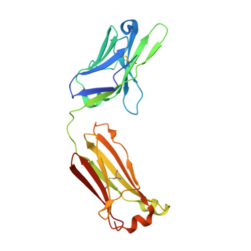
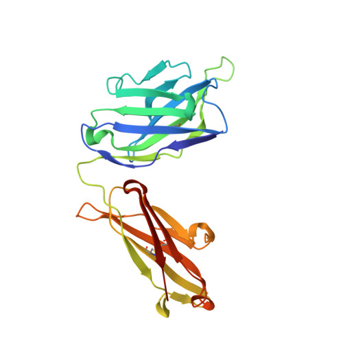
4GSD

H5.3 Fab Structure


Experimental Data Snapshot

  • Method: X-RAY DIFFRACTION
  • Resolution: 2.25 Å
  • R-Value Free: 0.203 
  • R-Value Work: 0.172 
  • R-Value Observed: 0.174 

Starting Model: experimental
View more details

wwPDB Validation   3D Report Full Report


This is version 1.4 of the entry. See complete history


Literature

Human antibodies that neutralize respiratory droplet transmissible H5N1 influenza viruses.

Thornburg, N.J.Nannemann, D.P.Blum, D.L.Belser, J.A.Tumpey, T.M.Deshpande, S.Fritz, G.A.Sapparapu, G.Krause, J.C.Lee, J.H.Ward, A.B.Lee, D.E.Li, S.Winarski, K.L.Spiller, B.W.Meiler, J.Crowe, J.E.

(2013) J Clin Invest 123: 4405-4409

  • DOI: https://doi.org/10.1172/JCI69377
  • Primary Citation of Related Structures:  
    4GSD

  • PubMed Abstract: 

    Recent studies described the experimental adaptation of influenza H5 HAs that confers respiratory droplet transmission (rdt) to influenza virus in ferrets. Acquisition of the ability to transmit via aerosol may lead to the development of a highly pathogenic pandemic H5 virus. Vaccines are predicted to play an important role in H5N1 control should the virus become readily transmissible between humans. We obtained PBMCs from patients who received an A/Vietnam/1203/2004 H5N1 subunit vaccine. Human hybridomas were then generated and characterized. We identified antibodies that bound the HA head domain and recognized both WT and rdt H5 HAs. We used a combination of structural techniques to define a mechanism of antibody recognition of an H5 HA receptor-binding site that neutralized H5N1 influenza viruses and pseudoviruses carrying the HA rdt variants that have mutations near the receptor-binding site. Incorporation or retention of this critical antigenic site should be considered in the design of novel H5 HA immunogens to protect against mammalian-adapted H5N1 mutants.


Macromolecules
Find similar proteins by:  (by identity cutoff)  |  3D Structure
Entity ID: 1
MoleculeChains Sequence LengthOrganismDetailsImage
H5.3 Fab Light ChainA [auth L]212Homo sapiensMutation(s): 0 
Entity Groups  
Sequence Clusters30% Identity50% Identity70% Identity90% Identity95% Identity100% Identity
Sequence Annotations
Expand
  • Reference Sequence
Find similar proteins by:  (by identity cutoff)  |  3D Structure
Entity ID: 2
MoleculeChains Sequence LengthOrganismDetailsImage
H5.3 Fab Heavy ChainB [auth H]226Homo sapiensMutation(s): 0 
Entity Groups  
Sequence Clusters30% Identity50% Identity70% Identity90% Identity95% Identity100% Identity
Sequence Annotations
Expand
  • Reference Sequence
Experimental Data & Validation

Experimental Data

  • Method: X-RAY DIFFRACTION
  • Resolution: 2.25 Å
  • R-Value Free: 0.203 
  • R-Value Work: 0.172 
  • R-Value Observed: 0.174 
  • Space Group: P 61
Unit Cell:
Length ( Å )Angle ( ˚ )
a = 150.43α = 90
b = 150.43β = 90
c = 70.517γ = 120
Software Package:
Software NamePurpose
MOLREPphasing
PHENIXrefinement
HKL-2000data reduction
HKL-2000data scaling

Structure Validation

View Full Validation Report


Entry History 

Deposition Data

Revision History  (Full details and data files)

  • Version 1.0: 2013-08-21
    Type: Initial release
  • Version 1.1: 2013-10-09
    Changes: Database references
  • Version 1.2: 2013-10-23
    Changes: Database references
  • Version 1.3: 2018-01-24
    Changes: Structure summary
  • Version 1.4: 2024-04-03
    Changes: Data collection, Database references, Refinement description