4GW3

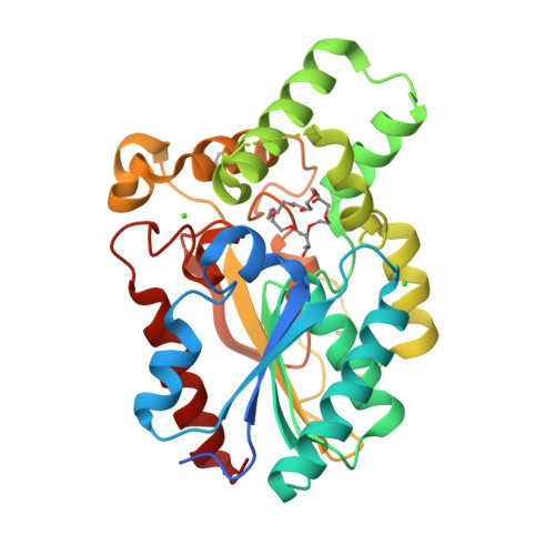
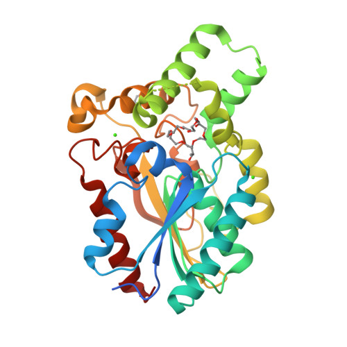
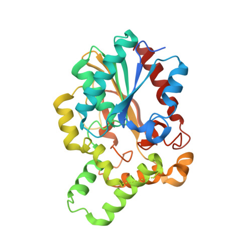
Crystal Structure of the Lipase from Proteus mirabilis


Experimental Data Snapshot

  • Method: X-RAY DIFFRACTION
  • Resolution: 2.00 Å
  • R-Value Free: 0.194 
  • R-Value Work: 0.167 
  • R-Value Observed: 0.169 

wwPDB Validation   3D Report Full Report


This is version 1.1 of the entry. See complete history


Literature

Crystal Structure of Proteus mirabilis Lipase, a Novel Lipase from the Proteus/Psychrophilic Subfamily of Lipase Family I.1.

Korman, T.P.Bowie, J.U.

(2012) PLoS One 7: e52890-e52890

  • DOI: https://doi.org/10.1371/journal.pone.0052890
  • Primary Citation of Related Structures:  
    4GW3, 4GXN

  • PubMed Abstract: 

    Bacterial lipases from family I.1 and I.2 catalyze the hydrolysis of triacylglycerol between 25-45°C and are used extensively as biocatalysts. The lipase from Proteus mirabilis belongs to the Proteus/psychrophilic subfamily of lipase family I.1 and is a promising catalyst for biodiesel production because it can tolerate high amounts of water in the reaction. Here we present the crystal structure of the Proteus mirabilis lipase, a member of the Proteus/psychrophilic subfamily of I.1lipases. The structure of the Proteus mirabilis lipase was solved in the absence and presence of a bound phosphonate inhibitor. Unexpectedly, both the apo and inhibitor bound forms of P. mirabilis lipase were found to be in a closed conformation. The structure reveals a unique oxyanion hole and a wide active site that is solvent accessible even in the closed conformation. A distinct mechanism for Ca²⁺ coordination may explain how these lipases can fold without specific chaperones.


  • Organizational Affiliation

    Department of Chemistry and Biochemisty, University of California Los Angeles, Los Angeles, California, United States of America.


Macromolecules
Find similar proteins by:  (by identity cutoff)  |  3D Structure
Entity ID: 1
MoleculeChains Sequence LengthOrganismDetailsImage
Putative lipase307Proteus mirabilisMutation(s): 0 
Gene Names: LipA
EC: 3.1.1.3
UniProt
Find proteins for B4EVM3 (Proteus mirabilis (strain HI4320))
Explore B4EVM3 
Go to UniProtKB:  B4EVM3
Entity Groups  
Sequence Clusters30% Identity50% Identity70% Identity90% Identity95% Identity100% Identity
UniProt GroupB4EVM3
Sequence Annotations
Expand
  • Reference Sequence
Small Molecules
Ligands 4 Unique
IDChains Name / Formula / InChI Key2D Diagram3D Interactions
1PE
Query on 1PE

Download Ideal Coordinates CCD File 
F [auth A],
G [auth A]
PENTAETHYLENE GLYCOL
C10 H22 O6
JLFNLZLINWHATN-UHFFFAOYSA-N
GOL
Query on GOL

Download Ideal Coordinates CCD File 
E [auth A]GLYCEROL
C3 H8 O3
PEDCQBHIVMGVHV-UHFFFAOYSA-N
IPA
Query on IPA

Download Ideal Coordinates CCD File 
D [auth A]ISOPROPYL ALCOHOL
C3 H8 O
KFZMGEQAYNKOFK-UHFFFAOYSA-N
CA
Query on CA

Download Ideal Coordinates CCD File 
B [auth A],
C [auth A]
CALCIUM ION
Ca
BHPQYMZQTOCNFJ-UHFFFAOYSA-N
Experimental Data & Validation

Experimental Data

  • Method: X-RAY DIFFRACTION
  • Resolution: 2.00 Å
  • R-Value Free: 0.194 
  • R-Value Work: 0.167 
  • R-Value Observed: 0.169 
  • Space Group: P 32
Unit Cell:
Length ( Å )Angle ( ˚ )
a = 65.438α = 90
b = 65.438β = 90
c = 63.966γ = 120
Software Package:
Software NamePurpose
SCALEPACKdata scaling
PHASERphasing
REFMACrefinement
PDB_EXTRACTdata extraction
CrystalCleardata collection
DENZOdata reduction

Structure Validation

View Full Validation Report


Entry History 

Deposition Data

  • Released Date: 2013-02-06 
  • Deposition Author(s): Korman, T.P.

Revision History  (Full details and data files)

  • Version 1.0: 2013-02-06
    Type: Initial release
  • Version 1.1: 2024-02-28
    Changes: Data collection, Database references, Derived calculations