


Crystal structure of glucose 1-phosphate thymidylyltransferase from Aneurinibacillus thermoaerophilus complexed with UDP-galactose and UTP
Chen, T.J., Chien, W.T., Lin, C.C., Wang, W.C.To be published.
Experimental Data Snapshot
Starting Model: experimental
View more details
Entity ID: 1 | |||||
|---|---|---|---|---|---|
| Molecule | Chains | Sequence Length | Organism | Details | Image |
| Glucose-1-phosphate thymidylyltransferase | 297 | Aneurinibacillus thermoaerophilus | Mutation(s): 0 Gene Names: rmlA EC: 2.7.7.24 | 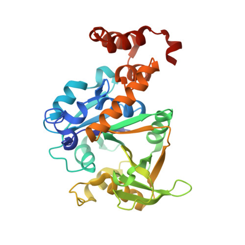 | |
UniProt | |||||
Find proteins for Q9AGY4 (Aneurinibacillus thermoaerophilus) Explore Q9AGY4 Go to UniProtKB: Q9AGY4 | |||||
Entity Groups | |||||
| Sequence Clusters | 30% Identity50% Identity70% Identity90% Identity95% Identity100% Identity | ||||
| UniProt Group | Q9AGY4 | ||||
Sequence AnnotationsExpand | |||||
| |||||
| Ligands 3 Unique | |||||
|---|---|---|---|---|---|
| ID | Chains | Name / Formula / InChI Key | 2D Diagram | 3D Interactions | |
| GDU Query on GDU | C [auth A], J [auth B] | GALACTOSE-URIDINE-5'-DIPHOSPHATE C15 H24 N2 O17 P2 HSCJRCZFDFQWRP-ABVWGUQPSA-N |  | ||
| UTP Query on UTP | D [auth A], K [auth B] | URIDINE 5'-TRIPHOSPHATE C9 H15 N2 O15 P3 PGAVKCOVUIYSFO-XVFCMESISA-N |  | ||
| SO4 Query on SO4 | E [auth A] F [auth A] G [auth A] H [auth A] I [auth A] | SULFATE ION O4 S QAOWNCQODCNURD-UHFFFAOYSA-L |  | ||
| Length ( Å ) | Angle ( ˚ ) |
|---|---|
| a = 114.788 | α = 90 |
| b = 90.763 | β = 125.2 |
| c = 87.702 | γ = 90 |
| Software Name | Purpose |
|---|---|
| DENZO | data reduction |
| SCALEPACK | data scaling |
| REFMAC | refinement |
| PDB_EXTRACT | data extraction |
| HKL-2000 | data collection |
| HKL-2000 | data reduction |
| MOLREP | phasing |
RCSB PDB is hosted by

RCSB PDB is a member of the


