4HSX

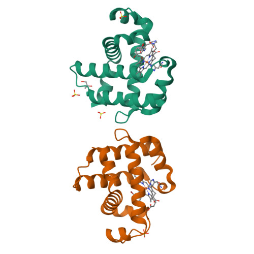
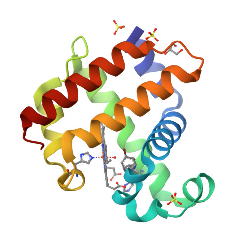
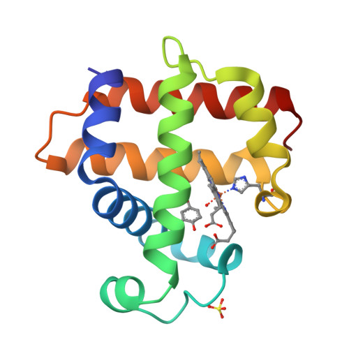
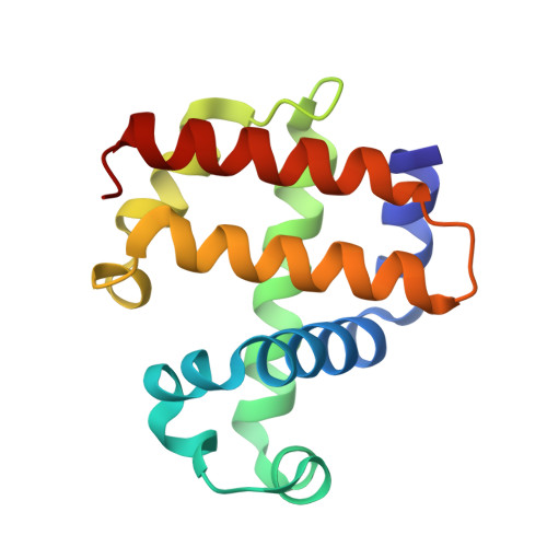
Structure of the L100F mutant of dehaloperoxidase-hemoglobin A from Amphitrite ornata with 4-bromophenol


Experimental Data Snapshot

  • Method: X-RAY DIFFRACTION
  • Resolution: 1.12 Å
  • R-Value Free: 0.150 
  • R-Value Work: 0.126 
  • R-Value Observed: 0.127 

wwPDB Validation   3D Report Full Report


Ligand Structure Quality Assessment 

Created with Raphaël 2.3.0Worse 01 BetterLigand structure goodness of fit to experimental dataBest fitted HEMClick on this verticalbar to view detailsBest fitted BMLClick on this verticalbar to view details

This is version 1.2 of the entry. See complete history


Literature

Role of polarity of the distal pocket in the control of inhibitor binding in dehaloperoxidase-hemoglobin.

Plummer, A.Thompson, M.K.Franzen, S.

(2013) Biochemistry 52: 2218-2227

  • DOI: https://doi.org/10.1021/bi301509r
  • Primary Citation of Related Structures:  
    4HSW, 4HSX

  • PubMed Abstract: 

    Dehaloperoxidase (DHP A), a unique multifunctional enzyme, from the marine annelid Amphitrite ornata dehalogenates 2,4,6-tribromophenol to form 2,6-dibromo-1,4-benzoquinone. The catalytic cycle of DHP is similar to that of horseradish peroxidase (HRP), involving a high-valent ferryl heme and two single-electron transfers from the aromatic substrate to the enzyme. Like HRP, DHP has been investigated as a potential bioremediation enzyme. However, DHP fails as a bioremediation enzyme because, unlike HRP, it has an internal binding cavity on the distal side of the heme capable of accommodating p-bromophenols, which act as an inhibitor of peroxidase function. Blocking internal binding in DHP may be the key to allowing the enzyme to function effectively as a peroxidase on the full range of halogenated phenols. The distal cavity of DHP is surrounded by several hydrophobic amino acids that stabilize internal binding of the monohalogenated phenols, including a leucine residue near the back edge of the heme (L100). We have expressed the L100F, L100Q, L100T, and L100V mutants of DHP in an effort to prevent internal binding and thereby convert the inhibitors into substrates. Kinetic assays and resonance Raman indicate that the peroxidase activity of the L100T and L100F mutants is increased compared to that of native DHP in the presence of 4-bromophenol (4-BP), suggesting a reduction in the inhibitor binding constant. In addition, the X-ray crystal structure of L100F clearly indicates a reduced occupancy of the 4-BP inhibitor in the distal cavity of DHP. However, at the same time, the L100F structure reveals that steric interference alone is insufficient to exclude the inhibitor. Instead, the comparison of L100T and isosteric L100V reveals that an increase in polarity plays a decisive role in excluding the inhibitor from the distal binding pocket.


  • Organizational Affiliation

    Department of Chemistry, North Carolina State University, Raleigh, NC 27695, USA.


Macromolecules
Find similar proteins by:  (by identity cutoff)  |  3D Structure
Entity ID: 1
MoleculeChains Sequence LengthOrganismDetailsImage
Dehaloperoxidase A
A, B
137Amphitrite ornataMutation(s): 1 
EC: 1.11.1.7
UniProt
Find proteins for Q9NAV8 (Amphitrite ornata)
Explore Q9NAV8 
Go to UniProtKB:  Q9NAV8
Entity Groups  
Sequence Clusters30% Identity50% Identity70% Identity90% Identity95% Identity100% Identity
UniProt GroupQ9NAV8
Sequence Annotations
Expand
  • Reference Sequence
Small Molecules
Ligands 4 Unique
IDChains Name / Formula / InChI Key2D Diagram3D Interactions
HEM
Query on HEM

Download Ideal Coordinates CCD File 
C [auth A],
I [auth B]
PROTOPORPHYRIN IX CONTAINING FE
C34 H32 Fe N4 O4
KABFMIBPWCXCRK-RGGAHWMASA-L
BML
Query on BML

Download Ideal Coordinates CCD File 
H [auth A],
K [auth B]
4-BROMOPHENOL
C6 H5 Br O
GZFGOTFRPZRKDS-UHFFFAOYSA-N
SO4
Query on SO4

Download Ideal Coordinates CCD File 
D [auth A],
E [auth A],
F [auth A],
J [auth B]
SULFATE ION
O4 S
QAOWNCQODCNURD-UHFFFAOYSA-L
GOL
Query on GOL

Download Ideal Coordinates CCD File 
G [auth A]GLYCEROL
C3 H8 O3
PEDCQBHIVMGVHV-UHFFFAOYSA-N
Experimental Data & Validation

Experimental Data

  • Method: X-RAY DIFFRACTION
  • Resolution: 1.12 Å
  • R-Value Free: 0.150 
  • R-Value Work: 0.126 
  • R-Value Observed: 0.127 
  • Space Group: P 21 21 21
Unit Cell:
Length ( Å )Angle ( ˚ )
a = 58.241α = 90
b = 67.441β = 90
c = 69.338γ = 90
Software Package:
Software NamePurpose
SCALEPACKdata scaling
REFMACrefinement
PDB_EXTRACTdata extraction
HKL-2000data collection
DENZOdata reduction
PHASERphasing

Structure Validation

View Full Validation Report


Ligand Structure Quality Assessment 

Created with Raphaël 2.3.0Worse 01 BetterLigand structure goodness of fit to experimental dataBest fitted HEMClick on this verticalbar to view detailsBest fitted BMLClick on this verticalbar to view details

Entry History 

Deposition Data

Revision History  (Full details and data files)

  • Version 1.0: 2013-05-01
    Type: Initial release
  • Version 1.1: 2017-11-15
    Changes: Refinement description
  • Version 1.2: 2024-02-28
    Changes: Data collection, Database references, Derived calculations