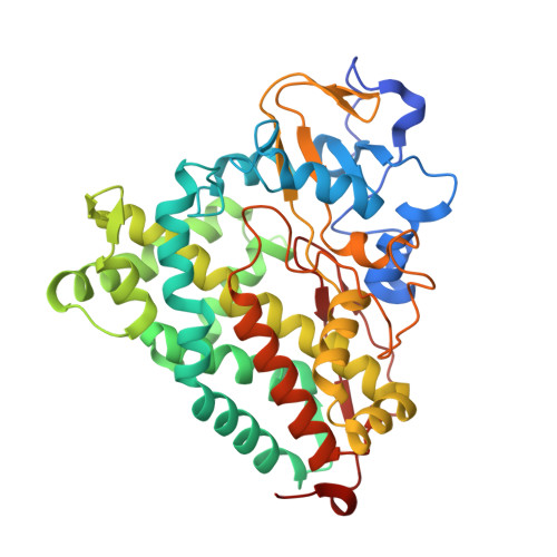
4L4G

Structure of cyanide and camphor bound P450cam mutant L358P/K178G


Experimental Data Snapshot

  • Method: X-RAY DIFFRACTION
  • Resolution: 1.55 Å
  • R-Value Free: 0.212 
  • R-Value Work: 0.172 
  • R-Value Observed: 0.174 

Starting Model: experimental
View more details

wwPDB Validation   3D Report Full Report


Ligand Structure Quality Assessment 


This is version 1.3 of the entry. See complete history


Literature

Synergistic Effects of Mutations in Cytochrome P450cam Designed To Mimic CYP101D1.

Batabyal, D.Li, H.Poulos, T.L.

(2013) Biochemistry 52: 5396-5402

  • DOI: https://doi.org/10.1021/bi400676d
  • Primary Citation of Related Structures:  
    4L49, 4L4A, 4L4B, 4L4C, 4L4D, 4L4E, 4L4F, 4L4G

  • PubMed Abstract: 

    A close orthologue to cytochrome P450cam (CYP101A1) that catalyzes the same hydroxylation of camphor to 5-exo-hydroxycamphor is CYP101D1. There are potentially important differences in and around the active site that could contribute to subtle functional differences. Adjacent to the heme iron ligand, Cys357, is Leu358 in P450cam, whereas this residue is Ala in CYP101D1. Leu358 plays a role in binding of the P450cam redox partner, putidaredoxin (Pdx). On the opposite side of the heme, about 15-20 Å away, Asp251 in P450cam plays a critical role in a proton relay network required for O2 activation but forms strong ion pairs with Arg186 and Lys178. In CYP101D1 Gly replaces Lys178. Thus, the local electrostatic environment and ion pairing are substantially different in CYP101D1. These sites have been systematically mutated in P450cam to the corresponding residues in CYP101D1 and the mutants analyzed by crystallography, kinetics, and UV-vis spectroscopy. Individually, the mutants have little effect on activity or structure, but in combination there is a major drop in enzyme activity. This loss in activity is due to the mutants being locked in the low-spin state, which prevents electron transfer from the P450cam redox partner, Pdx. These studies illustrate the strong synergistic effects on well-separated parts of the structure in controlling the equilibrium between the open (low-spin) and closed (high-spin) conformational states.


  • Organizational Affiliation

    Departments of Molecular Biology and Biochemistry, Chemistry, and Pharmaceutical Sciences, University of California, Irvine, CA 92697-3900, USA.


Macromolecules
Find similar proteins by:  (by identity cutoff)  |  3D Structure
Entity ID: 1
MoleculeChains Sequence LengthOrganismDetailsImage
Camphor 5-monooxygenase415Pseudomonas putidaMutation(s): 3 
Gene Names: camCcyp101
EC: 1.14.15.1
UniProt
Find proteins for P00183 (Pseudomonas putida)
Explore P00183 
Go to UniProtKB:  P00183
Entity Groups  
Sequence Clusters30% Identity50% Identity70% Identity90% Identity95% Identity100% Identity
UniProt GroupP00183
Sequence Annotations
Expand
  • Reference Sequence
Small Molecules
Ligands 4 Unique
IDChains Name / Formula / InChI Key2D Diagram3D Interactions
HEM
Query on HEM

Download Ideal Coordinates CCD File 
B [auth A]PROTOPORPHYRIN IX CONTAINING FE
C34 H32 Fe N4 O4
KABFMIBPWCXCRK-RGGAHWMASA-L
CAM
Query on CAM

Download Ideal Coordinates CCD File 
D [auth A]CAMPHOR
C10 H16 O
DSSYKIVIOFKYAU-XCBNKYQSSA-N
K
Query on K

Download Ideal Coordinates CCD File 
E [auth A],
F [auth A],
G [auth A]
POTASSIUM ION
K
NPYPAHLBTDXSSS-UHFFFAOYSA-N
CYN
Query on CYN

Download Ideal Coordinates CCD File 
C [auth A]CYANIDE ION
C N
XFXPMWWXUTWYJX-UHFFFAOYSA-N
Experimental Data & Validation

Experimental Data

  • Method: X-RAY DIFFRACTION
  • Resolution: 1.55 Å
  • R-Value Free: 0.212 
  • R-Value Work: 0.172 
  • R-Value Observed: 0.174 
  • Space Group: P 1 21 1
Unit Cell:
Length ( Å )Angle ( ˚ )
a = 57.072α = 90
b = 58.79β = 105.02
c = 57.072γ = 90
Software Package:
Software NamePurpose
Blu-Icedata collection
PHASERphasing
PHENIXrefinement
MOSFLMdata reduction
SCALAdata scaling

Structure Validation

View Full Validation Report


Ligand Structure Quality Assessment 


Entry History 

Deposition Data

Revision History  (Full details and data files)

  • Version 1.0: 2013-07-31
    Type: Initial release
  • Version 1.1: 2013-08-28
    Changes: Database references
  • Version 1.2: 2017-11-15
    Changes: Refinement description
  • Version 1.3: 2023-09-20
    Changes: Data collection, Database references, Derived calculations, Refinement description