4MZQ

beta-Alanyl-CoA:Ammonia Lyase from Clostridium propionicum in complex with propionyl-CoA


Experimental Data Snapshot

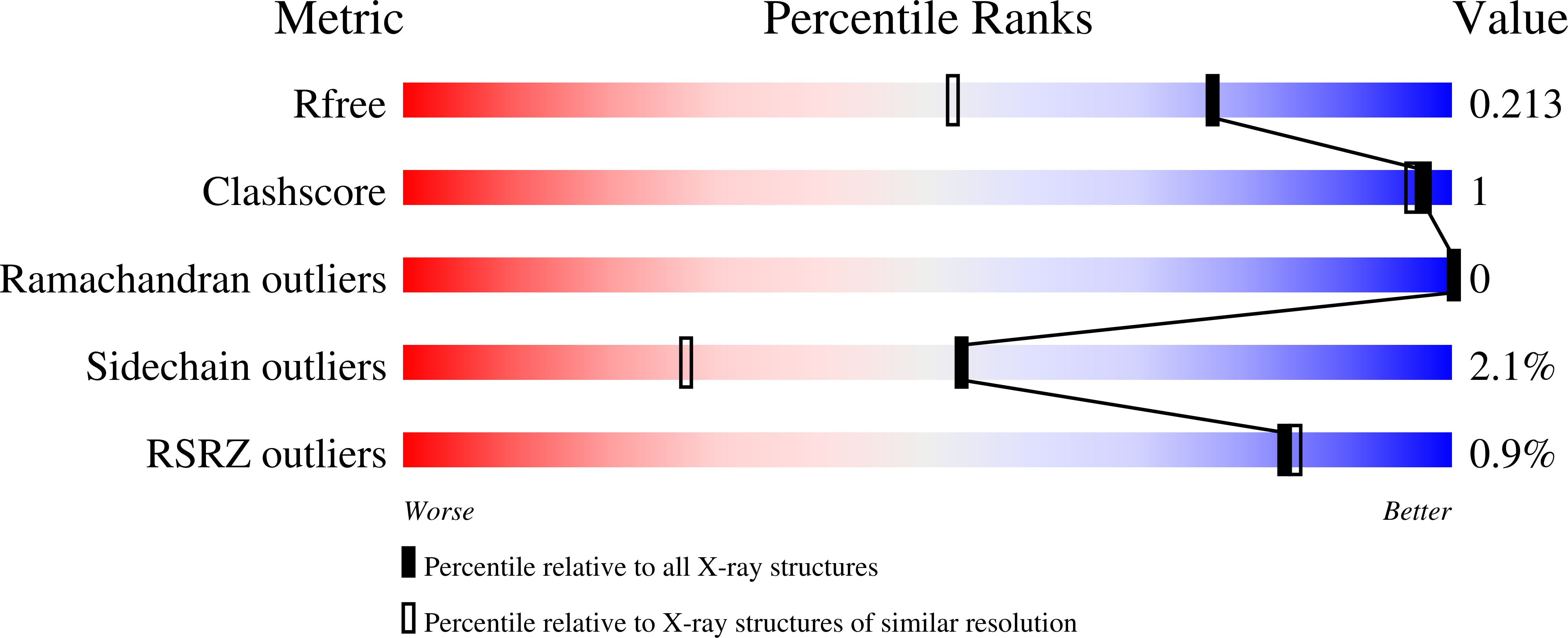
  • Method: X-RAY DIFFRACTION
  • Resolution: 1.59 Å
  • R-Value Free: 0.213 
  • R-Value Work: 0.179 
  • R-Value Observed: 0.181 

Starting Model: experimental
View more details

wwPDB Validation   3D Report Full Report


Ligand Structure Quality Assessment 


This is version 1.3 of the entry. See complete history


Literature

High resolution crystal structure of Clostridium propionicum beta-alanyl-CoA:ammonia lyase, a new member of the "hot dog fold" protein superfamily.

Heine, A.Herrmann, G.Selmer, T.Terwesten, F.Buckel, W.Reuter, K.

(2014) Proteins 82: 2041-2053

  • DOI: https://doi.org/10.1002/prot.24557
  • Primary Citation of Related Structures:  
    4MTU, 4MZQ

  • PubMed Abstract: 

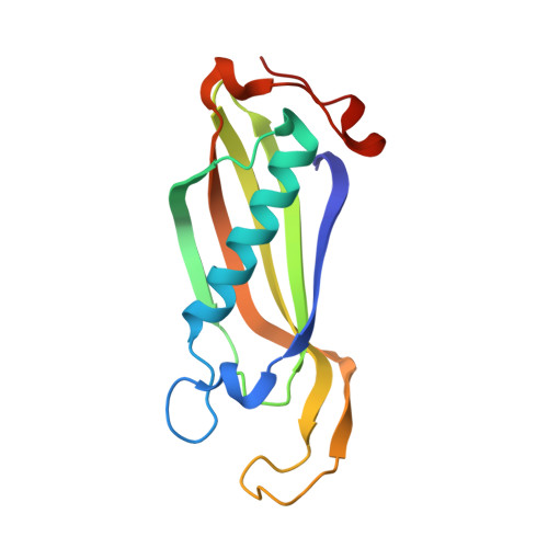
    Clostridium propionicum is the only organism known to ferment β-alanine, a constituent of coenzyme A (CoA) and the phosphopantetheinyl prosthetic group of holo-acyl carrier protein. The first step in the fermentation is a CoA-transfer to β-alanine. Subsequently, the resulting β-alanyl-CoA is deaminated by the enzyme β-alanyl-CoA:ammonia lyase (Acl) to reversibly form ammonia and acrylyl-CoA. We have determined the crystal structure of Acl in its apo-form at a resolution of 0.97 Å as well as in complex with CoA at a resolution of 1.59 Å. The structures reveal that the enyzme belongs to a superfamily of proteins exhibiting a so called "hot dog fold" which is characterized by a five-stranded antiparallel β-sheet with a long α-helix packed against it. The functional unit of all "hot dog fold" proteins is a homodimer containing two equivalent substrate binding sites which are established by the dimer interface. In the case of Acl, three functional dimers combine to a homohexamer strongly resembling the homohexamer formed by YciA-like acyl-CoA thioesterases. Here, we propose an enzymatic mechanism based on the crystal structure of the Acl·CoA complex and molecular docking.


  • Organizational Affiliation

    Institut für Pharmazeutische Chemie, Fachbereich Pharmazie, Philipps-Universität Marburg, Marbacher Weg 6, D-35032, Marburg, Germany.


Macromolecules
Find similar proteins by:  (by identity cutoff)  |  3D Structure
Entity ID: 1
MoleculeChains Sequence LengthOrganismDetailsImage
beta-Alanyl-CoA:Ammonia Lyase
A, B, C, D, E
A, B, C, D, E, F, G, H, I, J, K, L
144Anaerotignum propionicumMutation(s): 0 
UniProt
Find proteins for Q6KC22 (Anaerotignum propionicum)
Explore Q6KC22 
Go to UniProtKB:  Q6KC22
Entity Groups  
Sequence Clusters30% Identity50% Identity70% Identity90% Identity95% Identity100% Identity
UniProt GroupQ6KC22
Sequence Annotations
Expand
  • Reference Sequence
Small Molecules
Ligands 1 Unique
IDChains Name / Formula / InChI Key2D Diagram3D Interactions
1VU
Query on 1VU

Download Ideal Coordinates CCD File 
M [auth A]
N [auth B]
O [auth C]
P [auth D]
Q [auth E]
M [auth A],
N [auth B],
O [auth C],
P [auth D],
Q [auth E],
R [auth F],
S [auth G],
T [auth H],
U [auth I],
V [auth J],
W [auth K],
X [auth L]
propionyl Coenzyme A
C24 H40 N7 O17 P3 S
QAQREVBBADEHPA-IEXPHMLFSA-N
Experimental Data & Validation

Experimental Data

  • Method: X-RAY DIFFRACTION
  • Resolution: 1.59 Å
  • R-Value Free: 0.213 
  • R-Value Work: 0.179 
  • R-Value Observed: 0.181 
  • Space Group: P 1 21 1
Unit Cell:
Length ( Å )Angle ( ˚ )
a = 66.52α = 90
b = 110.965β = 89.98
c = 115.37γ = 90
Software Package:
Software NamePurpose
MAR345dtbdata collection
PHASERphasing
PHENIXrefinement
HKL-2000data reduction
HKL-2000data scaling

Structure Validation

View Full Validation Report


Ligand Structure Quality Assessment 


Entry History 

Deposition Data

Revision History  (Full details and data files)

  • Version 1.0: 2014-03-26
    Type: Initial release
  • Version 1.1: 2014-06-11
    Changes: Database references
  • Version 1.2: 2014-09-03
    Changes: Database references
  • Version 1.3: 2023-09-20
    Changes: Data collection, Database references, Derived calculations, Refinement description