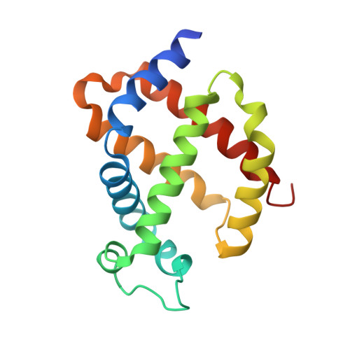
Crystallographic Studies with Xenon and Nitrous Oxide Provide Evidence for Protein-dependent Processes in the Mechanisms of General Anesthesia
Abraini, J.H., Marassio, G., David, H.N., Vallone, B., Prange, T., Colloc'h, N.(2014) Anesthesiology 121: 1018-1027
- PubMed: 25211169
- DOI: https://doi.org/10.1097/ALN.0000000000000435
- Primary Citation of Related Structures:
4NWE, 4NWH, 4NXA, 4NXC, 4O4T, 4O4Z - PubMed Abstract:
The mechanisms by which general anesthetics, including xenon and nitrous oxide, act are only beginning to be discovered. However, structural approaches revealed weak but specific protein-gas interactions. To improve knowledge, we performed x-ray crystallography studies under xenon and nitrous oxide pressure in a series of 10 binding sites within four proteins. Whatever the pressure, we show (1) hydrophobicity of the gas binding sites has a screening effect on xenon and nitrous oxide binding, with a threshold value of 83% beyond which and below which xenon and nitrous oxide, respectively, binds to their sites preferentially compared to each other; (2) xenon and nitrous oxide occupancies are significantly correlated respectively to the product and the ratio of hydrophobicity by volume, indicating that hydrophobicity and volume are binding parameters that complement and oppose each other's effects; and (3) the ratio of occupancy of xenon to nitrous oxide is significantly correlated to hydrophobicity of their binding sites. These data demonstrate that xenon and nitrous oxide obey different binding mechanisms, a finding that argues against all unitary hypotheses of narcosis and anesthesia, and indicate that the Meyer-Overton rule of a high correlation between anesthetic potency and solubility in lipids of general anesthetics is often overinterpreted. This study provides evidence that the mechanisms of gas binding to proteins and therefore of general anesthesia should be considered as the result of a fully reversible interaction between a drug ligand and a receptor as this occurs in classical pharmacology.
Organizational Affiliation:
From the Faculté de Médecine, Université de Caen Basse Normandie, Normandie-Université, Caen, France (J.H.A., G.M.); Institut de Recherche Biomédicale des Armées, Brétigny-sur-Orge, France (J.H.A.); Département d'Anesthésiologie, Université Laval, Quebec City, Québec, Canada (J.H.A., H.N.D.); Centre de Recherche Hôtel-Dieu de Lévis, CSSS Alphonse-Desjardins, Lévis, Québec, Canada (H.N.D.); Department of Biochemical Sciences, University of Rome La Sapienza, Rome, Italy (B.V.); LCRB UMR 8015, Université Paris Descartes-CNRS, Faculté de Pharmacie, Paris, France (T.P.); ISTCT UMR 6301, CNRS, CERVOxy group, GIP Cyceron, Caen, France (N.C.); ISTCT UMR 6301, Université de Caen Basse Normandie, Normandie-Université, Caen, France (N.C.); and ISTCT UMR 6301, CEA DSV/I2BM, Caen, France (N.C.).


