4POV

ThiT with LMG135 bound


Experimental Data Snapshot

  • Method: X-RAY DIFFRACTION
  • Resolution: 2.20 Å
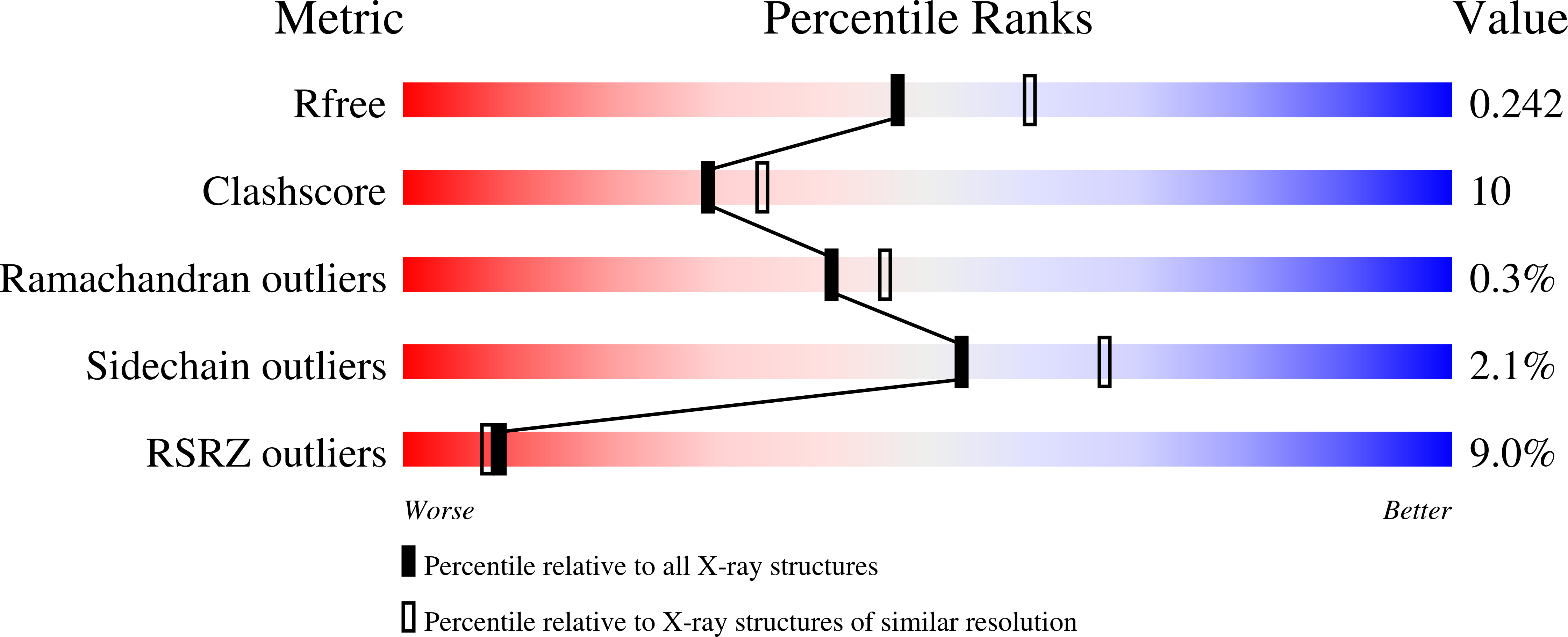
  • R-Value Free: 0.237 
  • R-Value Work: 0.185 
  • R-Value Observed: 0.185 

Starting Model: experimental
View more details

wwPDB Validation   3D Report Full Report


Ligand Structure Quality Assessment 


This is version 1.3 of the entry. See complete history


Literature

Structure-Based Design of Potent Small-Molecule Binders to the S-Component of the ECF Transporter for Thiamine.

Swier, L.J.Monjas, L.Guskov, A.de Voogd, A.R.Erkens, G.B.Slotboom, D.J.Hirsch, A.K.

(2015) Chembiochem 16: 819-826

  • DOI: https://doi.org/10.1002/cbic.201402673
  • Primary Citation of Related Structures:  
    4POP, 4POV

  • PubMed Abstract: 

    Energy-coupling factor (ECF) transporters are membrane-protein complexes that mediate vitamin uptake in prokaryotes. They bind the substrate through the action of a specific integral membrane subunit (S-component) and power transport by hydrolysis of ATP in the three-subunit ECF module. Here, we have studied the binding of thiamine derivatives to ThiT, a thiamine-specific S-component. We designed and synthesized derivatives of thiamine that bind to ThiT with high affinity; this allowed us to evaluate the contribution of the functional groups to the binding affinity. We determined six crystal structures of ThiT in complex with our derivatives. The structure of the substrate-binding site in ThiT remains almost unchanged despite substantial differences in affinity. This work indicates that the structural organization of the binding site is robust and suggests that substrate release, which is required for transport, requires additional changes in conformation in ThiT that might be imposed by the ECF module.


  • Organizational Affiliation

    Groningen Biomolecular Sciences and Biotechnology Institute, University of Groningen, Nijenborgh 4, 9747 AG Groningen (The Netherlands) http://www.rug.nl/research/membrane-enzymology/


Macromolecules
Find similar proteins by:  (by identity cutoff)  |  3D Structure
Entity ID: 1
MoleculeChains Sequence LengthOrganismDetailsImage
Thiamine transporter ThiT
A, B
192Lactococcus cremoris subsp. cremoris MG1363Mutation(s): 0 
Gene Names: llmg_0334LLNZ_01755thiT
Membrane Entity: Yes 
UniProt
Find proteins for A2RI47 (Lactococcus lactis subsp. cremoris (strain MG1363))
Explore A2RI47 
Go to UniProtKB:  A2RI47
Entity Groups  
Sequence Clusters30% Identity50% Identity70% Identity90% Identity95% Identity100% Identity
UniProt GroupA2RI47
Sequence Annotations
Expand
  • Reference Sequence
Small Molecules
Ligands 9 Unique
IDChains Name / Formula / InChI Key2D Diagram3D Interactions
BNG
Query on BNG

Download Ideal Coordinates CCD File 
CA [auth B]
DA [auth B]
EA [auth B]
J [auth A]
K [auth A]
CA [auth B],
DA [auth B],
EA [auth B],
J [auth A],
K [auth A],
L [auth A]
nonyl beta-D-glucopyranoside
C15 H30 O6
QFAPUKLCALRPLH-UXXRCYHCSA-N
P6G
Query on P6G

Download Ideal Coordinates CCD File 
V [auth A]HEXAETHYLENE GLYCOL
C12 H26 O7
IIRDTKBZINWQAW-UHFFFAOYSA-N
2VY
Query on 2VY

Download Ideal Coordinates CCD File 
C [auth A],
W [auth B]
4-[(4-amino-2-methylpyrimidin-5-yl)methyl]-3-methylthiophene-2-carbaldehyde
C12 H13 N3 O S
NWGVSSQLYKZANM-UHFFFAOYSA-N
1PE
Query on 1PE

Download Ideal Coordinates CCD File 
KA [auth B],
LA [auth B],
R [auth A]
PENTAETHYLENE GLYCOL
C10 H22 O6
JLFNLZLINWHATN-UHFFFAOYSA-N
PG4
Query on PG4

Download Ideal Coordinates CCD File 
AA [auth B]
BA [auth B]
E [auth A]
F [auth A]
G [auth A]
AA [auth B],
BA [auth B],
E [auth A],
F [auth A],
G [auth A],
H [auth A],
I [auth A],
Y [auth B],
Z [auth B]
TETRAETHYLENE GLYCOL
C8 H18 O5
UWHCKJMYHZGTIT-UHFFFAOYSA-N
PGE
Query on PGE

Download Ideal Coordinates CCD File 
S [auth A],
T [auth A],
U [auth A]
TRIETHYLENE GLYCOL
C6 H14 O4
ZIBGPFATKBEMQZ-UHFFFAOYSA-N
PG0
Query on PG0

Download Ideal Coordinates CCD File 
D [auth A],
X [auth B]
2-(2-METHOXYETHOXY)ETHANOL
C5 H12 O3
SBASXUCJHJRPEV-UHFFFAOYSA-N
PEG
Query on PEG

Download Ideal Coordinates CCD File 
FA [auth B]
GA [auth B]
HA [auth B]
IA [auth B]
JA [auth B]
FA [auth B],
GA [auth B],
HA [auth B],
IA [auth B],
JA [auth B],
M [auth A],
N [auth A],
O [auth A],
P [auth A],
Q [auth A]
DI(HYDROXYETHYL)ETHER
C4 H10 O3
MTHSVFCYNBDYFN-UHFFFAOYSA-N
CL
Query on CL

Download Ideal Coordinates CCD File 
MA [auth B]CHLORIDE ION
Cl
VEXZGXHMUGYJMC-UHFFFAOYSA-M
Binding Affinity Annotations 
IDSourceBinding Affinity
2VY BindingDB:  4POV Kd: 7.44 (nM) from 1 assay(s)
Experimental Data & Validation

Experimental Data

  • Method: X-RAY DIFFRACTION
  • Resolution: 2.20 Å
  • R-Value Free: 0.237 
  • R-Value Work: 0.185 
  • R-Value Observed: 0.185 
  • Space Group: C 1 2 1
Unit Cell:
Length ( Å )Angle ( ˚ )
a = 62.72α = 90
b = 84.35β = 96.08
c = 127.25γ = 90
Software Package:
Software NamePurpose
MxCuBEdata collection
CCP4model building
PHENIXrefinement
XDSdata reduction
XSCALEdata scaling
CCP4phasing

Structure Validation

View Full Validation Report


Ligand Structure Quality Assessment 


Entry History 

Deposition Data

Revision History  (Full details and data files)

  • Version 1.0: 2014-09-17
    Type: Initial release
  • Version 1.1: 2015-04-01
    Changes: Database references
  • Version 1.2: 2020-07-29
    Type: Remediation
    Reason: Carbohydrate remediation
    Changes: Data collection, Database references, Derived calculations, Structure summary
  • Version 1.3: 2023-09-20
    Changes: Data collection, Database references, Refinement description, Structure summary