4Q2T | pdb_00004q2t

Crystal structure of Arginyl-tRNA synthetase complexed with L-arginine


Experimental Data Snapshot

  • Method: X-RAY DIFFRACTION
  • Resolution: 2.40 Å
  • R-Value Free: 
    0.258 (Depositor), 0.260 (DCC) 
  • R-Value Work: 
    0.196 (Depositor), 0.200 (DCC) 
  • R-Value Observed: 
    0.198 (Depositor) 

Starting Model: experimental
View more details

wwPDB Validation   3D Report Full Report


Ligand Structure Quality Assessment 

Created with Raphaël 2.3.0Worse 01 BetterLigand structure goodness of fit to experimental dataBest fitted ARGClick on this verticalbar to view details

This is version 1.2 of the entry. See complete history


Literature

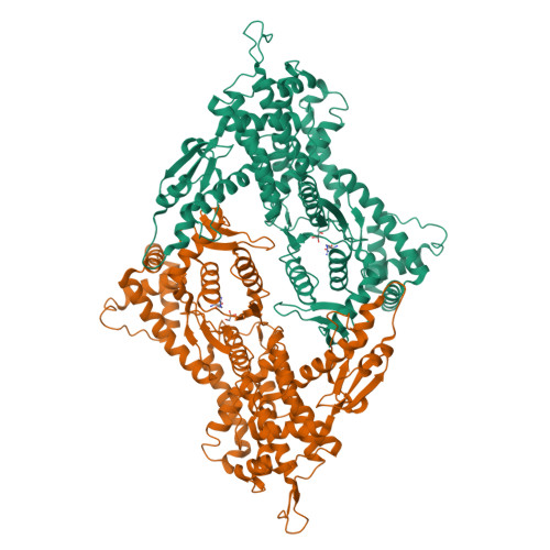
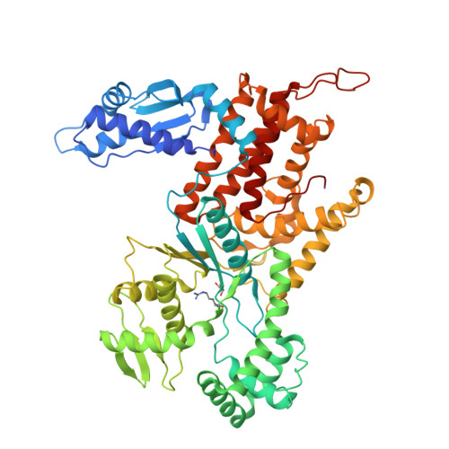
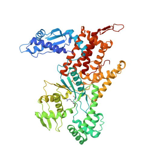
The crystal structure of arginyl-tRNA synthetase from Homo sapiens

Kim, H.S.Cha, S.Y.Jo, C.H.Han, A.R.Hwang, K.Y.

(2014) FEBS Lett 588: 2328-2334

  • DOI: https://doi.org/10.1016/j.febslet.2014.05.027
  • Primary Citation of Related Structures:  
    4Q2T, 4Q2X, 4Q2Y

  • PubMed Abstract: 

    Arginyl-tRNA synthetase (ArgRS) is a tRNA-binding protein that catalyzes the esterification of L-arginine to its cognate tRNA. L-Canavanine, a structural analog of L-arginine, has recently been studied as an anticancer agent. Here, we determined the crystal structures of the apo, L-arginine-complexed, and L-canavanine-complexed forms of the cytoplasmic free isoform of human ArgRS (hArgRS). Similar interactions were formed upon binding to L-canavanine or L-arginine, but the interaction between Tyr312 and the oxygen of the oxyguanidino group was a little bit different. Detailed conformational changes that occur upon substrate binding were explained. The hArgRS structure was also compared with previously reported homologue structures. The results presented here may provide a basis for the design of new anticancer drugs, such as L-canavanine analogs.


  • Organizational Affiliation

    Division of Biotechnology, College of Life Sciences and Biotechnology, Korea University, Seoul, Republic of Korea.


Macromolecules
Find similar proteins by:  (by identity cutoff)  |  3D Structure
Entity ID: 1
MoleculeChains Sequence LengthOrganismDetailsImage
Arginine--tRNA ligase, cytoplasmic
A, B
607Homo sapiensMutation(s): 1 
Gene Names: RARS
EC: 6.1.1.19
UniProt & NIH Common Fund Data Resources
Find proteins for P54136 (Homo sapiens)
Explore P54136 
Go to UniProtKB:  P54136
PHAROS:  P54136
GTEx:  ENSG00000113643 
Entity Groups  
Sequence Clusters30% Identity50% Identity70% Identity90% Identity95% Identity100% Identity
UniProt GroupP54136
Sequence Annotations
Expand
  • Reference Sequence
Experimental Data & Validation

Experimental Data

  • Method: X-RAY DIFFRACTION
  • Resolution: 2.40 Å
  • R-Value Free:  0.258 (Depositor), 0.260 (DCC) 
  • R-Value Work:  0.196 (Depositor), 0.200 (DCC) 
  • R-Value Observed: 0.198 (Depositor) 
Space Group: P 21 21 21
Unit Cell:
Length ( Å )Angle ( ˚ )
a = 74.485α = 90
b = 106.901β = 90
c = 175.248γ = 90
Software Package:
Software NamePurpose
DENZOdata reduction
SCALEPACKdata scaling
PHENIXrefinement
PDB_EXTRACTdata extraction
HKL-2000data collection
PHASERphasing

Structure Validation

View Full Validation Report


Ligand Structure Quality Assessment 

Created with Raphaël 2.3.0Worse 01 BetterLigand structure goodness of fit to experimental dataBest fitted ARGClick on this verticalbar to view details

Entry History 

Deposition Data

Revision History  (Full details and data files)

  • Version 1.0: 2014-07-23
    Type: Initial release
  • Version 1.1: 2017-11-22
    Changes: Refinement description
  • Version 1.2: 2023-11-08
    Changes: Data collection, Database references, Derived calculations, Refinement description