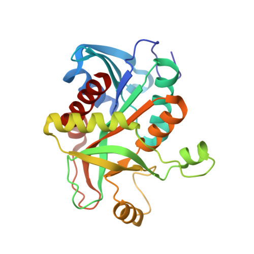
4TS9

Crystal structure of wild type E. Coli purine nucleoside phosphorylase with 6 FMC molecules


Experimental Data Snapshot

  • Method: X-RAY DIFFRACTION
  • Resolution: 1.77 Å
  • R-Value Free: 0.177 
  • R-Value Work: 0.156 
  • R-Value Observed: 0.157 

Starting Model: experimental
View more details

wwPDB Validation   3D Report Full Report


Ligand Structure Quality Assessment 


This is version 1.3 of the entry. See complete history


Literature

Crystallographic snapshots of ligand binding to hexameric purine nucleoside phosphorylase and kinetic studies give insight into the mechanism of catalysis.

Stefanic, Z.Narczyk, M.Mikleusevic, G.Kazazic, S.Bzowska, A.Luic, M.

(2018) Sci Rep 8: 15427-15427

  • DOI: https://doi.org/10.1038/s41598-018-33723-1
  • Primary Citation of Related Structures:  
    4TS3, 4TS9, 4TTA, 4TTI, 4TTJ

  • PubMed Abstract: 

    Purine nucleoside phosphorylase (PNP) catalyses the cleavage of the glycosidic bond of purine nucleosides using phosphate instead of water as a second substrate. PNP from Escherichia coli is a homohexamer, build as a trimer of dimers, and each subunit can be in two conformations, open or closed. This conformational change is induced by the presence of phosphate substrate, and very likely a required step for the catalysis. Closing one active site strongly affects the others, by a yet unclear mechanism and order of events. Kinetic and ligand binding studies show strong negative cooperativity between subunits. Here, for the first time, we managed to monitor the sequence of nucleoside binding to individual subunits in the crystal structures of the wild-type enzyme, showing that first the closed sites, not the open ones, are occupied by the nucleoside. However, two mutations within the active site, Asp204Ala/Arg217Ala, are enough not only to significantly reduce the effectiveness of the enzyme, but also reverse the sequence of the nucleoside binding. In the mutant the open sites, neighbours in a dimer of those in the closed conformation, are occupied as first. This demonstrates how important for the effective catalysis of Escherichia coli PNP is proper subunit cooperation.


  • Organizational Affiliation

    Division of Physical Chemistry, Ruđer Bošković Institute, Bijenička 54, Zagreb, 10000, Croatia.


Macromolecules
Find similar proteins by:  (by identity cutoff)  |  3D Structure
Entity ID: 1
MoleculeChains Sequence LengthOrganismDetailsImage
Purine nucleoside phosphorylase DeoD-type
A, B, C
237Escherichia coliMutation(s): 0 
EC: 2.4.2.1
UniProt
Find proteins for P0ABP8 (Escherichia coli (strain K12))
Explore P0ABP8 
Go to UniProtKB:  P0ABP8
Entity Groups  
Sequence Clusters30% Identity50% Identity70% Identity90% Identity95% Identity100% Identity
UniProt GroupP0ABP8
Sequence Annotations
Expand
  • Reference Sequence
Experimental Data & Validation

Experimental Data

  • Method: X-RAY DIFFRACTION
  • Resolution: 1.77 Å
  • R-Value Free: 0.177 
  • R-Value Work: 0.156 
  • R-Value Observed: 0.157 
  • Space Group: P 61 2 2
Unit Cell:
Length ( Å )Angle ( ˚ )
a = 120.625α = 90
b = 120.625β = 90
c = 240.006γ = 120
Software Package:
Software NamePurpose
XDSdata reduction
PDB_EXTRACTdata extraction
PHENIXrefinement
XSCALEdata scaling

Structure Validation

View Full Validation Report


Ligand Structure Quality Assessment 


Entry History 

Deposition Data

Revision History  (Full details and data files)

  • Version 1.0: 2015-07-08
    Type: Initial release
  • Version 1.1: 2016-03-16
    Changes: Database references, Structure summary
  • Version 1.2: 2018-10-31
    Changes: Data collection, Database references
  • Version 1.3: 2023-12-20
    Changes: Data collection, Database references, Refinement description