4U2L | pdb_00004u2l

Dithionite reduced cholesterol in complex with sulfite


Experimental Data Snapshot

  • Method: X-RAY DIFFRACTION
  • Resolution: 1.34 Å
  • R-Value Free: 
    0.133 (Depositor), 0.140 (DCC) 
  • R-Value Work: 
    0.097 (Depositor), 0.110 (DCC) 
  • R-Value Observed: 
    0.101 (Depositor) 

Starting Model: experimental
View more details

wwPDB Validation   3D Report Full Report


Ligand Structure Quality Assessment 

Created with Raphaël 2.3.0Worse 01 BetterLigand structure goodness of fit to experimental dataBest fitted SFDClick on this verticalbar to view details

This is version 1.5 of the entry. See complete history


Literature

High-resolution structures of cholesterol oxidase in the reduced state provide insights into redox stabilization.

Golden, E.Karton, A.Vrielink, A.

(2014) Acta Crystallogr D Biol Crystallogr 70: 3155-3166

  • DOI: https://doi.org/10.1107/S139900471402286X
  • Primary Citation of Related Structures:  
    4U2L, 4U2S, 4U2T

  • PubMed Abstract: 

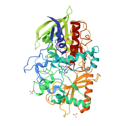
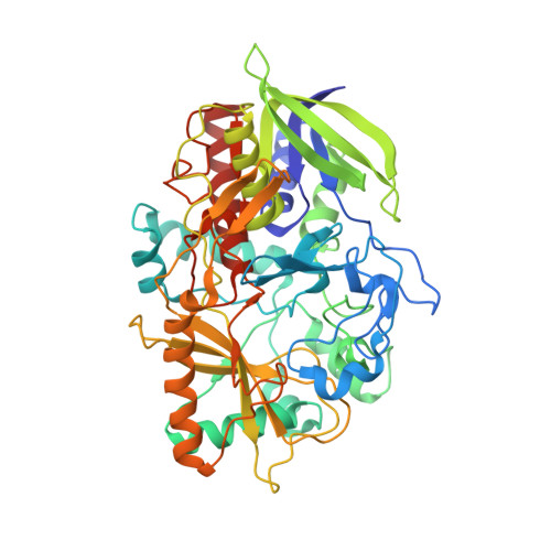
    Cholesterol oxidase (CO) is a flavoenzyme that catalyzes the oxidation and isomerization of cholesterol to cholest-4-en-3-one. The reductive half reaction occurs via a hydride transfer from the substrate to the FAD cofactor. The structures of CO reduced with dithionite under aerobic conditions and in the presence of the substrate 2-propanol under both aerobic and anaerobic conditions are presented. The 1.32 Å resolution structure of the dithionite-reduced enzyme reveals a sulfite molecule covalently bound to the FAD cofactor. The isoalloxazine ring system displays a bent structure relative to that of the oxidized enzyme, and alternate conformations of a triad of aromatic residues near to the cofactor are evident. A 1.12 Å resolution anaerobically trapped reduced enzyme structure in the presence of 2-propanol does not show a similar bending of the flavin ring system, but does show alternate conformations of the aromatic triad. Additionally, a significant difference electron-density peak is observed within a covalent-bond distance of N5 of the flavin moiety, suggesting that a hydride-transfer event has occurred as a result of substrate oxidation trapping the flavin in the electron-rich reduced state. The hydride transfer generates a tetrahedral geometry about the flavin N5 atom. High-level density-functional theory calculations were performed to correlate the crystallographic findings with the energetics of this unusual arrangement of the flavin moiety. These calculations suggest that strong hydrogen-bond interactions between Gly120 and the flavin N5 centre may play an important role in these structural features.


  • Organizational Affiliation

    School of Chemistry and Biochemistry, University of Western Australia, Crawley, Western Australia 6009, Australia.


Macromolecules
Find similar proteins by:  (by identity cutoff)  |  3D Structure
Entity ID: 1
MoleculeChains Sequence LengthOrganismDetailsImage
Cholesterol oxidase510Streptomyces sp. SA-COOMutation(s): 0 
Gene Names: choA
EC: 1.1.3.6 (PDB Primary Data), 5.3.3.1 (PDB Primary Data)
UniProt
Find proteins for P12676 (Streptomyces sp. (strain SA-COO))
Explore P12676 
Go to UniProtKB:  P12676
Entity Groups  
Sequence Clusters30% Identity50% Identity70% Identity90% Identity95% Identity100% Identity
UniProt GroupP12676
Sequence Annotations
Expand
  • Reference Sequence
Small Molecules
Ligands 2 Unique
IDChains Name / Formula / InChI Key2D Diagram3D Interactions
SFD
Query on SFD

Download Ideal Coordinates CCD File 
E [auth A](S)-10-((2S,3S,4R)-5-((S)-((S)-(((2R,3S,4R,5R)-5-(6-AMINO-9H-PURIN-9-YL)-3,4-DIHYDROXY-TETRAHYDROFURAN-2-YL)METHOXY)(HYDROXY)PHOSPHORYLOXY)(HYDROXY)PHOSPHORYLOXY)-2,3,4-TRIHYDROXYPENTYL)-7,8-DIMETHYL-2,4-DIOXO-2,3,4,4A-TETRAHYDROBENZO[G]PTERIDINE-5(10H)-SULFONIC ACID
C27 H35 N9 O18 P2 S
WCBOBURWKHIISA-LDVRFEIFSA-N
SO4
Query on SO4

Download Ideal Coordinates CCD File 
B [auth A],
C [auth A],
D [auth A]
SULFATE ION
O4 S
QAOWNCQODCNURD-UHFFFAOYSA-L
Experimental Data & Validation

Experimental Data

  • Method: X-RAY DIFFRACTION
  • Resolution: 1.34 Å
  • R-Value Free:  0.133 (Depositor), 0.140 (DCC) 
  • R-Value Work:  0.097 (Depositor), 0.110 (DCC) 
  • R-Value Observed: 0.101 (Depositor) 
Space Group: P 1 21 1
Unit Cell:
Length ( Å )Angle ( ˚ )
a = 51.375α = 90
b = 73.565β = 105.03
c = 63.431γ = 90
Software Package:
Software NamePurpose
PHENIXrefinement
SCALAdata scaling
PDB_EXTRACTdata extraction
PHASERphasing

Structure Validation

View Full Validation Report


Ligand Structure Quality Assessment 

Created with Raphaël 2.3.0Worse 01 BetterLigand structure goodness of fit to experimental dataBest fitted SFDClick on this verticalbar to view details

Entry History 

Deposition Data

Revision History  (Full details and data files)

  • Version 1.0: 2014-12-10
    Type: Initial release
  • Version 1.1: 2014-12-24
    Changes: Database references
  • Version 1.2: 2015-02-04
    Changes: Derived calculations
  • Version 1.3: 2016-06-01
    Changes: Data collection
  • Version 1.4: 2017-11-22
    Changes: Data collection, Derived calculations, Refinement description
  • Version 1.5: 2023-09-27
    Changes: Data collection, Database references, Refinement description