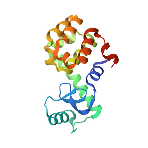
Homologous ligands accommodated by discrete conformations of a buried cavity.
Merski, M., Fischer, M., Balius, T.E., Eidam, O., Shoichet, B.K.(2015) Proc Natl Acad Sci U S A 112: 5039-5044
- PubMed: 25847998
- DOI: https://doi.org/10.1073/pnas.1500806112
- Primary Citation of Related Structures:
4W51, 4W52, 4W53, 4W54, 4W55, 4W56, 4W57, 4W58, 4W59 - PubMed Abstract:
Conformational change in protein-ligand complexes is widely modeled, but the protein accommodation expected on binding a congeneric series of ligands has received less attention. Given their use in medicinal chemistry, there are surprisingly few substantial series of congeneric ligand complexes in the Protein Data Bank (PDB). Here we determine the structures of eight alkyl benzenes, in single-methylene increases from benzene to n-hexylbenzene, bound to an enclosed cavity in T4 lysozyme. The volume of the apo cavity suffices to accommodate benzene but, even with toluene, larger cavity conformations become observable in the electron density, and over the series two other major conformations are observed. These involve discrete changes in main-chain conformation, expanding the site; few continuous changes in the site are observed. In most structures, two discrete protein conformations are observed simultaneously, and energetic considerations suggest that these conformations are low in energy relative to the ground state. An analysis of 121 lysozyme cavity structures in the PDB finds that these three conformations dominate the previously determined structures, largely modeled in a single conformation. An investigation of the few congeneric series in the PDB suggests that discrete changes are common adaptations to a series of growing ligands. The discrete, but relatively few, conformational states observed here, and their energetic accessibility, may have implications for anticipating protein conformational change in ligand design.
Organizational Affiliation:
Department of Pharmaceutical Chemistry, University of California, San Francisco, CA 94158-2550.


