4YJI

The Crystal Structure of a Bacterial Aryl Acylamidase Belonging to the Amidase signature (AS) enzymes family


Experimental Data Snapshot

  • Method: X-RAY DIFFRACTION
  • Resolution: 1.73 Å
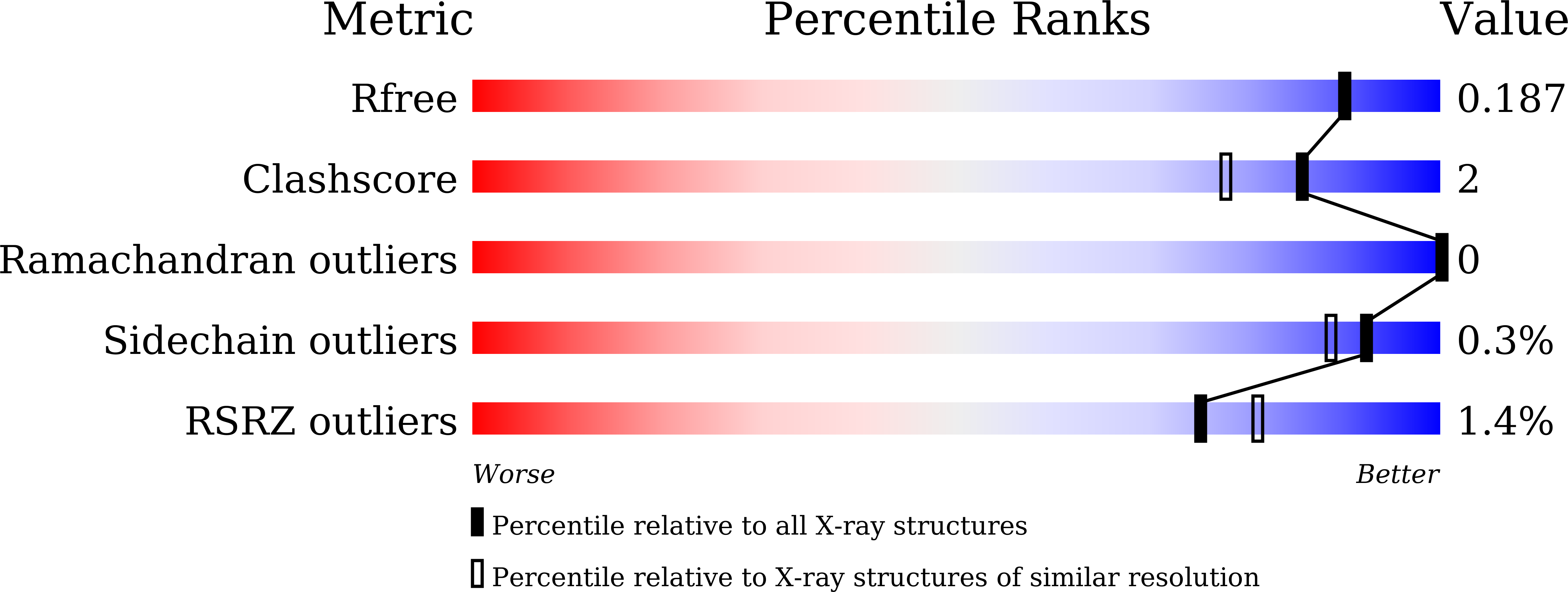
  • R-Value Free: 0.188 
  • R-Value Work: 0.167 
  • R-Value Observed: 0.167 

Starting Model: experimental
View more details

wwPDB Validation   3D Report Full Report


Ligand Structure Quality Assessment 


This is version 1.1 of the entry. See complete history


Literature

Crystal structure analysis of a bacterial aryl acylamidase belonging to the amidase signature enzyme family

Lee, S.Park, E.-H.Ko, H.-J.Bang, W.-G.Kim, H.Y.Kim, K.H.Choi, I.-G.

(2015) Biochem Biophys Res Commun 467: 268-274

  • DOI: https://doi.org/10.1016/j.bbrc.2015.09.177
  • Primary Citation of Related Structures:  
    4YJ6, 4YJI

  • PubMed Abstract: 

    The atomic structure of a bacterial aryl acylamidase (EC 3.5.1.13; AAA) is reported and structural features are investigated to better understand the catalytic profile of this enzyme. Structures of AAA were determined in its native form and in complex with the analgesic acetanilide, p-acetaminophenol, at 1.70 Å and 1.73 Å resolutions, respectively. The overall structural fold of AAA was identified as an α/β fold class, exhibiting an open twisted β-sheet core surrounded by α-helices. The asymmetric unit contains one AAA molecule and the monomeric form is functionally active. The core structure enclosing the signature sequence region, including the canonical Ser-cisSer-Lys catalytic triad, is conserved in all members of the Amidase Signature enzyme family. The structure of AAA in a complex with its ligand reveals a unique organization in the substrate-binding pocket. The binding pocket consists of two loops (loop1 and loop2) in the amidase signature sequence and one helix (α10) in the non-amidase signature sequence. We identified two residues (Tyr(136) and Thr(330)) that interact with the ligand via water molecules, and a hydrogen-bonding network that explains the catalytic affinity over various aryl acyl compounds. The optimum activity of AAA at pH > 10 suggests that the reaction mechanism employs Lys(84) as the catalytic base to polarize the Ser(187) nucleophile in the catalytic triad.


  • Organizational Affiliation

    Department of Biotechnology, College of Life Sciences and Biotechnology, Korea University, Anam-Dong, Seoungbuk-Gu, Seoul, 136-713, South Korea.


Macromolecules
Find similar proteins by:  (by identity cutoff)  |  3D Structure
Entity ID: 1
MoleculeChains Sequence LengthOrganismDetailsImage
Aryl acylamidase495bacterium CSBL00001Mutation(s): 1 
EC: 3.5.1.13
UniProt
Find proteins for C3UWD1 (bacterium CSBL00001)
Explore C3UWD1 
Go to UniProtKB:  C3UWD1
Entity Groups  
Sequence Clusters30% Identity50% Identity70% Identity90% Identity95% Identity100% Identity
UniProt GroupC3UWD1
Sequence Annotations
Expand
  • Reference Sequence
Small Molecules
Ligands 2 Unique
IDChains Name / Formula / InChI Key2D Diagram3D Interactions
CXS
Query on CXS

Download Ideal Coordinates CCD File 
B [auth A]3-CYCLOHEXYL-1-PROPYLSULFONIC ACID
C9 H19 N O3 S
PJWWRFATQTVXHA-UHFFFAOYSA-N
TYL
Query on TYL

Download Ideal Coordinates CCD File 
C [auth A]N-(4-HYDROXYPHENYL)ACETAMIDE (TYLENOL)
C8 H9 N O2
RZVAJINKPMORJF-UHFFFAOYSA-N
Experimental Data & Validation

Experimental Data

  • Method: X-RAY DIFFRACTION
  • Resolution: 1.73 Å
  • R-Value Free: 0.188 
  • R-Value Work: 0.167 
  • R-Value Observed: 0.167 
  • Space Group: P 41 21 2
Unit Cell:
Length ( Å )Angle ( ˚ )
a = 75.98α = 90
b = 75.98β = 90
c = 214.71γ = 90
Software Package:
Software NamePurpose
PHENIXrefinement
MOSFLMdata reduction
MOSFLMdata scaling
PHENIXphasing

Structure Validation

View Full Validation Report


Ligand Structure Quality Assessment 


Entry History 

Deposition Data

Revision History  (Full details and data files)

  • Version 1.0: 2015-11-04
    Type: Initial release
  • Version 1.1: 2023-11-08
    Changes: Data collection, Database references, Derived calculations, Refinement description