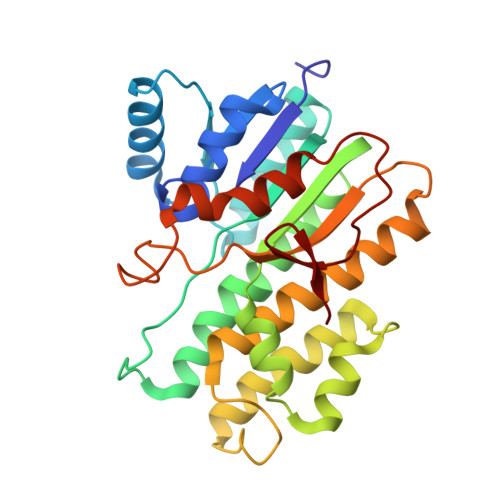
4Z3D

Human carbonyl reductase 1 with glutathione in a protective configuration


Experimental Data Snapshot

  • Method: X-RAY DIFFRACTION
  • Resolution: 1.80 Å
  • R-Value Free: 0.212 
  • R-Value Work: 0.182 
  • R-Value Observed: 0.183 

wwPDB Validation   3D Report Full Report


Ligand Structure Quality Assessment 


This is version 1.1 of the entry. See complete history


Literature

Structural insights on the catalytic site protection of human carbonyl reductase 1 by glutathione.

Liang, Q.Liu, R.Du, S.Ding, Y.

(2015) J Struct Biol 192: 138-144

  • DOI: https://doi.org/10.1016/j.jsb.2015.09.005
  • Primary Citation of Related Structures:  
    4Z3D

  • PubMed Abstract: 

    The NADPH-dependent human carbonyl reductase 1 (hCBR1), a member of the short-chain dehydrogenase/reductase protein family, plays an important role in the ubiquitous metabolism of endogenous and xenobiotic carbonyl containing compounds. Glutathione (GSH) is also a cofactor of hCBR1, however, its role in the carbonyl reductase function of the enzyme is still unclear. In this study, we presented the crystal structure of hCBR1 in complex with GSH, in the absence of its substrates or inhibitors. Interestingly, we found that the GSH molecule presents in a configuration quite different from that was previously reported when substrate is binding to hCBR1. Our structure indicates that GSH contributes to the substrate selectivity of hCBR1 and protects the catalytic center of hCBR1 through a switch-like mechanism. The isothermal titration calorimetry and enzymology data shows that GSH directly binding with hCBR1 when there's no substrate exist. The enzymology data also shows GSH protects NADPH being attacked by oxidative small molecules. This is the first time that GSH is found to demonstrate such functions as a co-enzyme. Our crystal structure succeeds in providing critical insights into the substrate selectivity of hCBR1 and the interaction between hCBR1 and GSH.


  • Organizational Affiliation

    Department of Physiology and Biophysics, School of Life Sciences, Fudan University, Shanghai 200438, China.


Macromolecules
Find similar proteins by:  (by identity cutoff)  |  3D Structure
Entity ID: 1
MoleculeChains Sequence LengthOrganismDetailsImage
Carbonyl reductase [NADPH] 1
A, B, C, D
276Homo sapiensMutation(s): 0 
Gene Names: CBR1CBRCRNSDR21C1
EC: 1.1.1.184 (PDB Primary Data), 1.1.1.197 (PDB Primary Data), 1.1.1.189 (PDB Primary Data), 1.1.1.196 (UniProt), 1.1.1.71 (UniProt)
UniProt & NIH Common Fund Data Resources
Find proteins for P16152 (Homo sapiens)
Explore P16152 
Go to UniProtKB:  P16152
PHAROS:  P16152
GTEx:  ENSG00000159228 
Entity Groups  
Sequence Clusters30% Identity50% Identity70% Identity90% Identity95% Identity100% Identity
UniProt GroupP16152
Sequence Annotations
Expand
  • Reference Sequence
Small Molecules
Ligands 2 Unique
IDChains Name / Formula / InChI Key2D Diagram3D Interactions
NDP
Query on NDP

Download Ideal Coordinates CCD File 
F [auth A],
H [auth B],
J [auth C],
L [auth D]
NADPH DIHYDRO-NICOTINAMIDE-ADENINE-DINUCLEOTIDE PHOSPHATE
C21 H30 N7 O17 P3
ACFIXJIJDZMPPO-NNYOXOHSSA-N
GSH
Query on GSH

Download Ideal Coordinates CCD File 
E [auth A],
G [auth B],
I [auth C],
K [auth D]
GLUTATHIONE
C10 H17 N3 O6 S
RWSXRVCMGQZWBV-WDSKDSINSA-N
Experimental Data & Validation

Experimental Data

  • Method: X-RAY DIFFRACTION
  • Resolution: 1.80 Å
  • R-Value Free: 0.212 
  • R-Value Work: 0.182 
  • R-Value Observed: 0.183 
  • Space Group: P 1
Unit Cell:
Length ( Å )Angle ( ˚ )
a = 55.567α = 82.99
b = 57.18β = 87.32
c = 99.453γ = 85.66
Software Package:
Software NamePurpose
PHENIXrefinement
HKL-2000data scaling
MOLREPphasing

Structure Validation

View Full Validation Report


Ligand Structure Quality Assessment 


Entry History 

Deposition Data

Revision History  (Full details and data files)

  • Version 1.0: 2015-10-14
    Type: Initial release
  • Version 1.1: 2024-03-20
    Changes: Data collection, Database references, Derived calculations