5CKR

Crystal Structure of MraY in complex with Muraymycin D2


Experimental Data Snapshot

  • Method: X-RAY DIFFRACTION
  • Resolution: 2.95 Å
  • R-Value Free: 0.261 
  • R-Value Work: 0.247 
  • R-Value Observed: 0.248 

Starting Model: experimental
View more details

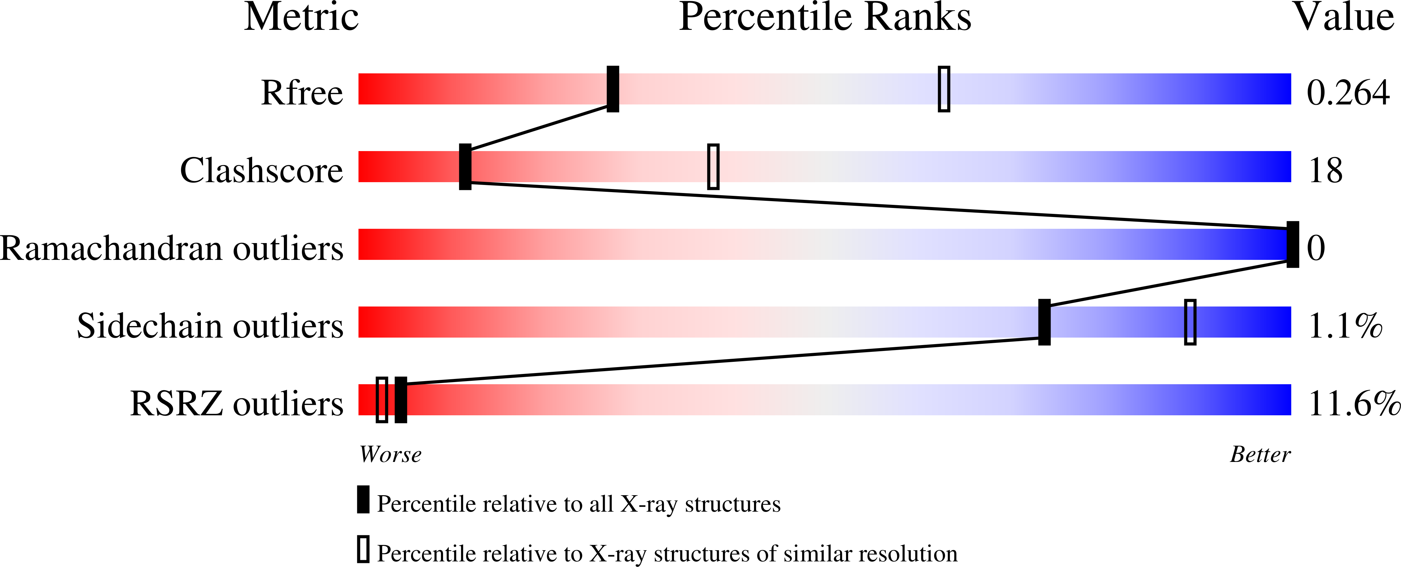
wwPDB Validation   3D Report Full Report


Ligand Structure Quality Assessment 


This is version 1.4 of the entry. See complete history


Literature

Structural insights into inhibition of lipid I production in bacterial cell wall synthesis.

Chung, B.C.Mashalidis, E.H.Tanino, T.Kim, M.Matsuda, A.Hong, J.Ichikawa, S.Lee, S.Y.

(2016) Nature 533: 557-560

  • DOI: https://doi.org/10.1038/nature17636
  • Primary Citation of Related Structures:  
    5CKR

  • PubMed Abstract: 

    Antibiotic-resistant bacterial infection is a serious threat to public health. Peptidoglycan biosynthesis is a well-established target for antibiotic development. MraY (phospho-MurNAc-pentapeptide translocase) catalyses the first and an essential membrane step of peptidoglycan biosynthesis. It is considered a very promising target for the development of new antibiotics, as many naturally occurring nucleoside inhibitors with antibacterial activity target this enzyme. However, antibiotics targeting MraY have not been developed for clinical use, mainly owing to a lack of structural insight into inhibition of this enzyme. Here we present the crystal structure of MraY from Aquifex aeolicus (MraYAA) in complex with its naturally occurring inhibitor, muraymycin D2 (MD2). We show that after binding MD2, MraYAA undergoes remarkably large conformational rearrangements near the active site, which lead to the formation of a nucleoside-binding pocket and a peptide-binding site. MD2 binds the nucleoside-binding pocket like a two-pronged plug inserting into a socket. Further interactions it makes in the adjacent peptide-binding site anchor MD2 to and enhance its affinity for MraYAA. Surprisingly, MD2 does not interact with three acidic residues or the Mg(2+) cofactor required for catalysis, suggesting that MD2 binds to MraYAA in a manner that overlaps with, but is distinct from, its natural substrate, UDP-MurNAc-pentapeptide. We have determined the principles of MD2 binding to MraYAA, including how it avoids the need for pyrophosphate and sugar moieties, which are essential features for substrate binding. The conformational plasticity of MraY could be the reason that it is the target of many structurally distinct inhibitors. These findings can inform the design of new inhibitors targeting MraY as well as its paralogues, WecA and TarO.


  • Organizational Affiliation

    Department of Biochemistry, Duke University Medical Center, 303 Research Drive, Durham, North Carolina, 27710, USA.


Macromolecules
Find similar proteins by:  (by identity cutoff)  |  3D Structure
Entity ID: 1
MoleculeChains Sequence LengthOrganismDetailsImage
Phospho-N-acetylmuramoyl-pentapeptide-transferase365Aquifex aeolicus VF5Mutation(s): 0 
Gene Names: mraYaq_053
EC: 2.7.8.13
Membrane Entity: Yes 
UniProt
Find proteins for O66465 (Aquifex aeolicus (strain VF5))
Explore O66465 
Go to UniProtKB:  O66465
Entity Groups  
Sequence Clusters30% Identity50% Identity70% Identity90% Identity95% Identity100% Identity
UniProt GroupO66465
Sequence Annotations
Expand
  • Reference Sequence
Small Molecules
Ligands 1 Unique
IDChains Name / Formula / InChI Key2D Diagram3D Interactions
57M
Query on 57M

Download Ideal Coordinates CCD File 
B [auth A]Muraymycin D2
C37 H61 N11 O16
RRTIONDZEJYWBN-VDXVSALRSA-N
Experimental Data & Validation

Experimental Data

  • Method: X-RAY DIFFRACTION
  • Resolution: 2.95 Å
  • R-Value Free: 0.261 
  • R-Value Work: 0.247 
  • R-Value Observed: 0.248 
  • Space Group: C 2 2 21
Unit Cell:
Length ( Å )Angle ( ˚ )
a = 94.48α = 90
b = 102.05β = 90
c = 135.8γ = 90
Software Package:
Software NamePurpose
PHENIXrefinement
MOSFLMdata reduction
Cootmodel building
iMOSFLMdata reduction
Aimlessdata scaling
PHASERphasing

Structure Validation

View Full Validation Report


Ligand Structure Quality Assessment 


Entry History & Funding Information

Deposition Data


Funding OrganizationLocationGrant Number
National Institutes of Health/National Institute of General Medical Sciences (NIH/NIGMS)United StatesR01 GM100984

Revision History  (Full details and data files)

  • Version 1.0: 2016-03-02
    Type: Initial release
  • Version 1.1: 2016-06-01
    Changes: Database references
  • Version 1.2: 2017-09-27
    Changes: Author supporting evidence, Database references, Derived calculations, Refinement description
  • Version 1.3: 2019-12-25
    Changes: Author supporting evidence
  • Version 1.4: 2023-09-27
    Changes: Data collection, Database references, Refinement description, Structure summary