5H7L | pdb_00005h7l

Complex of Elongation factor 2-50S ribosomal protein L12


Experimental Data Snapshot

  • Method: X-RAY DIFFRACTION
  • Resolution: 3.10 Å
  • R-Value Free: 
    0.285 (Depositor), 0.290 (DCC) 
  • R-Value Work: 
    0.230 (Depositor), 0.230 (DCC) 
  • R-Value Observed: 
    0.233 (Depositor) 

wwPDB Validation   3D Report Full Report


Ligand Structure Quality Assessment 

Created with Raphaël 2.3.0Worse 01 BetterLigand structure goodness of fit to experimental dataBest fitted GCPClick on this verticalbar to view details

This is version 1.3 of the entry. See complete history


Literature

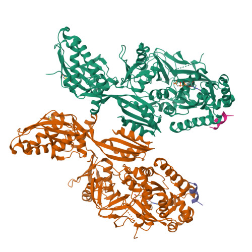
The C-terminal helix of ribosomal P stalk recognizes a hydrophobic groove of elongation factor 2 in a novel fashion

Tanzawa, T.Kato, K.Girodat, D.Ose, T.Kumakura, Y.Wieden, H.J.Uchiumi, T.Tanaka, I.Yao, M.

(2018) Nucleic Acids Res 46: 3232-3244

  • DOI: https://doi.org/10.1093/nar/gky115
  • Primary Citation of Related Structures:  
    5H7J, 5H7K, 5H7L

  • PubMed Abstract: 

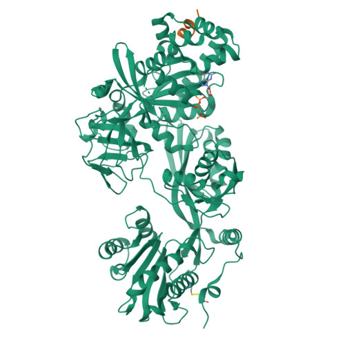
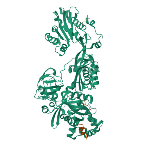
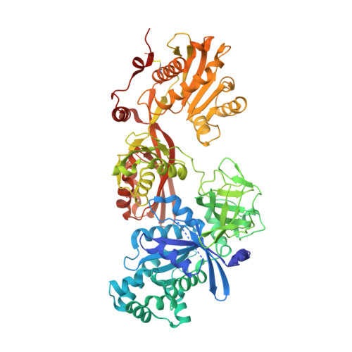
    Archaea and eukaryotes have ribosomal P stalks composed of anchor protein P0 and aP1 homodimers (archaea) or P1•P2 heterodimers (eukaryotes). These P stalks recruit translational GTPases to the GTPase-associated center in ribosomes to provide energy during translation. The C-terminus of the P stalk is known to selectively recognize GTPases. Here we investigated the interaction between the P stalk and elongation factor 2 by determining the structures of Pyrococcus horikoshii EF-2 (PhoEF-2) in the Apo-form, GDP-form, GMPPCP-form (GTP-form), and GMPPCP-form bound with 11 C-terminal residues of P1 (P1C11). Helical structured P1C11 binds to a hydrophobic groove between domain G and subdomain G' of PhoEF-2, where is completely different from that of aEF-1α in terms of both position and sequence, implying that such interaction characteristic may be requested by how GTPases perform their functions on the ribosome. Combining PhoEF-2 P1-binding assays with a structural comparison of current PhoEF-2 structures and molecular dynamics model of a P1C11-bound GDP form, the conformational changes of the P1C11-binding groove in each form suggest that in response to the translation process, the groove has three states: closed, open, and release for recruiting and releasing GTPases.


  • Organizational Affiliation

    Graduate School of Life Science, Hokkaido University, Sapporo, Hokkaido 060-0810, Japan.


Macromolecules
Find similar proteins by:  (by identity cutoff)  |  3D Structure
Entity ID: 1
MoleculeChains Sequence LengthOrganismDetailsImage
Elongation factor 2
A, B
743Pyrococcus horikoshii OT3Mutation(s): 0 
Gene Names: fusA
UniProt
Find proteins for O59521 (Pyrococcus horikoshii (strain ATCC 700860 / DSM 12428 / JCM 9974 / NBRC 100139 / OT-3))
Explore O59521 
Go to UniProtKB:  O59521
Entity Groups  
Sequence Clusters30% Identity50% Identity70% Identity90% Identity95% Identity100% Identity
UniProt GroupO59521
Sequence Annotations
Expand
  • Reference Sequence

Find similar proteins by:  Sequence   |   3D Structure  

Entity ID: 2
MoleculeChains Sequence LengthOrganismDetailsImage
50S ribosomal protein L12C [auth G],
D [auth E]
11Pyrococcus horikoshii OT3Mutation(s): 0 
UniProt
Find proteins for O57705 (Pyrococcus horikoshii (strain ATCC 700860 / DSM 12428 / JCM 9974 / NBRC 100139 / OT-3))
Explore O57705 
Go to UniProtKB:  O57705
Entity Groups  
Sequence Clusters30% Identity50% Identity70% Identity90% Identity95% Identity100% Identity
UniProt GroupO57705
Sequence Annotations
Expand
  • Reference Sequence
Experimental Data & Validation

Experimental Data

  • Method: X-RAY DIFFRACTION
  • Resolution: 3.10 Å
  • R-Value Free:  0.285 (Depositor), 0.290 (DCC) 
  • R-Value Work:  0.230 (Depositor), 0.230 (DCC) 
  • R-Value Observed: 0.233 (Depositor) 
Space Group: P 21 21 21
Unit Cell:
Length ( Å )Angle ( ˚ )
a = 79.774α = 90
b = 121.902β = 90
c = 199.265γ = 90
Software Package:
Software NamePurpose
PHENIXrefinement
XDSdata reduction
XDSdata scaling
MOLREPphasing

Structure Validation

View Full Validation Report


Ligand Structure Quality Assessment 

Created with Raphaël 2.3.0Worse 01 BetterLigand structure goodness of fit to experimental dataBest fitted GCPClick on this verticalbar to view details

Entry History & Funding Information

Deposition Data


Funding OrganizationLocationGrant Number
Japan Society for the Promotion of ScienceJapan26650013
Japan Society for the Promoting of ScienceJapan24370073

Revision History  (Full details and data files)

  • Version 1.0: 2018-02-21
    Type: Initial release
  • Version 1.1: 2018-03-07
    Changes: Database references
  • Version 1.2: 2018-05-02
    Changes: Data collection, Database references
  • Version 1.3: 2024-10-16
    Changes: Data collection, Database references, Structure summary