5JSG | pdb_00005jsg

Crystal structure of Spindlin1 bound to compound EML405


Experimental Data Snapshot

  • Method: X-RAY DIFFRACTION
  • Resolution: 2.50 Å
  • R-Value Free: 
    0.245 (Depositor), 0.240 (DCC) 
  • R-Value Work: 
    0.186 (Depositor), 0.190 (DCC) 
  • R-Value Observed: 
    0.192 (Depositor) 

wwPDB Validation   3D Report Full Report


Ligand Structure Quality Assessment 

Created with Raphaël 2.3.0Worse 01 BetterLigand structure goodness of fit to experimental dataBest fitted 6P9Click on this verticalbar to view details

This is version 1.2 of the entry. See complete history


Literature

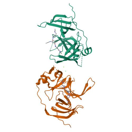
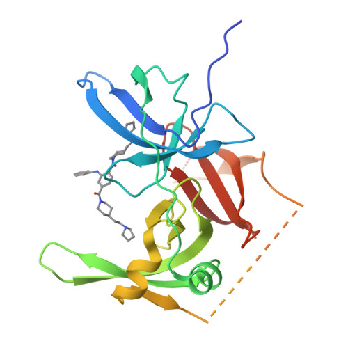
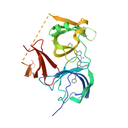
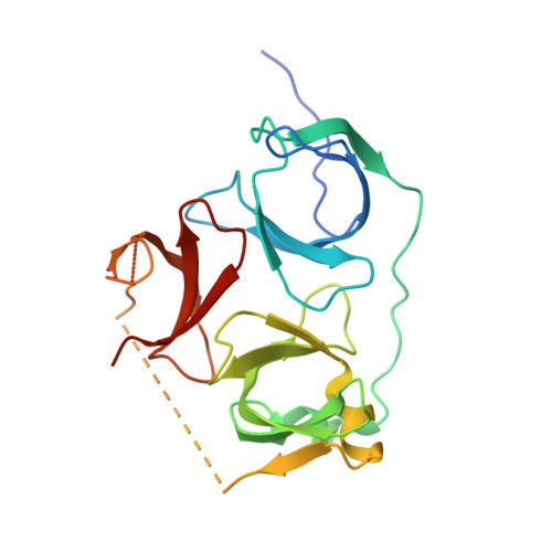
Developing Spindlin1 small-molecule inhibitors by using protein microarrays

Bae, N.Viviano, M.Su, X.Lv, J.Cheng, D.Sagum, C.Castellano, S.Bai, X.Johnson, C.Khalil, M.I.Shen, J.Chen, K.Li, H.Sbardella, G.Bedford, M.T.

(2017) Nat Chem Biol 13: 750-756

  • DOI: https://doi.org/10.1038/nchembio.2377
  • Primary Citation of Related Structures:  
    5JSG, 5JSJ

  • PubMed Abstract: 

    The discovery of inhibitors of methyl- and acetyl-binding domains has provided evidence for the 'druggability' of epigenetic effector molecules. The small-molecule probe UNC1215 prevents methyl-dependent protein-protein interactions by engaging the aromatic cage of MBT domains and, with lower affinity, Tudor domains. Using a library of tagged UNC1215 analogs, we screened a protein-domain microarray of human methyllysine effector molecules to rapidly detect compounds with new binding profiles with either increased or decreased specificity. Using this approach, we identified a compound (EML405) that acquired a novel interaction with the Tudor-domain-containing protein Spindlin1 (SPIN1). Structural studies facilitated the rational synthesis of SPIN1 inhibitors with increased selectivity (EML631-633), which engage SPIN1 in cells, block its ability to 'read' H3K4me3 marks and inhibit its transcriptional-coactivator activity. Protein microarrays can thus be used as a platform to 'target-hop' and identify small molecules that bind and compete with domain-motif interactions.


  • Organizational Affiliation

    Department of Epigenetics and Molecular Carcinogenesis, University of Texas MD Anderson Cancer Center, Smithville, Texas, USA.


Macromolecules
Find similar proteins by:  (by identity cutoff)  |  3D Structure
Entity ID: 1
MoleculeChains Sequence LengthOrganismDetailsImage
Spindlin-1
A, B
222Homo sapiensMutation(s): 0 
Gene Names: SPIN1OCRSPIN
UniProt & NIH Common Fund Data Resources
Find proteins for Q9Y657 (Homo sapiens)
Explore Q9Y657 
Go to UniProtKB:  Q9Y657
PHAROS:  Q9Y657
GTEx:  ENSG00000106723 
Entity Groups  
Sequence Clusters30% Identity50% Identity70% Identity90% Identity95% Identity100% Identity
UniProt GroupQ9Y657
Sequence Annotations
Expand
  • Reference Sequence
Experimental Data & Validation

Experimental Data

  • Method: X-RAY DIFFRACTION
  • Resolution: 2.50 Å
  • R-Value Free:  0.245 (Depositor), 0.240 (DCC) 
  • R-Value Work:  0.186 (Depositor), 0.190 (DCC) 
  • R-Value Observed: 0.192 (Depositor) 
Space Group: P 1 21 1
Unit Cell:
Length ( Å )Angle ( ˚ )
a = 43.06α = 90
b = 123.908β = 92.3
c = 49.779γ = 90
Software Package:
Software NamePurpose
HKL-2000data collection
HKL-2000data scaling
PHENIXrefinement
PDB_EXTRACTdata extraction
HKL-2000data reduction
MOLREPphasing

Structure Validation

View Full Validation Report


Ligand Structure Quality Assessment 

Created with Raphaël 2.3.0Worse 01 BetterLigand structure goodness of fit to experimental dataBest fitted 6P9Click on this verticalbar to view details

Entry History 

Deposition Data

  • Released Date: 2017-05-10 
  • Deposition Author(s): Su, X., Li, H.

Revision History  (Full details and data files)

  • Version 1.0: 2017-05-10
    Type: Initial release
  • Version 1.1: 2017-07-05
    Changes: Database references
  • Version 1.2: 2024-03-20
    Changes: Data collection, Database references, Derived calculations