5UV8 | pdb_00005uv8

Interleukin-3 Receptor Complex


Experimental Data Snapshot

  • Method: X-RAY DIFFRACTION
  • Resolution: 2.70 Å
  • R-Value Free: 
    0.286 (Depositor), 0.290 (DCC) 
  • R-Value Work: 
    0.242 (Depositor), 0.240 (DCC) 
  • R-Value Observed: 
    0.244 (Depositor) 

Starting Models: experimental
View more details

wwPDB Validation   3D Report Full Report


Ligand Structure Quality Assessment 

Created with Raphaël 2.3.0Worse 01 BetterLigand structure goodness of fit to experimental dataBest fitted NAGClick on this verticalbar to view details

This is version 2.2 of the entry. See complete history


Literature

A dual role for the N-terminal domain of the IL-3 receptor in cell signalling.

Broughton, S.E.Hercus, T.R.Nero, T.L.Kan, W.L.Barry, E.F.Dottore, M.Cheung Tung Shing, K.S.Morton, C.J.Dhagat, U.Hardy, M.P.Wilson, N.J.Downton, M.T.Schieber, C.Hughes, T.P.Lopez, A.F.Parker, M.W.

(2018) Nat Commun 9: 386-386

  • DOI: https://doi.org/10.1038/s41467-017-02633-7
  • Primary Citation of Related Structures:  
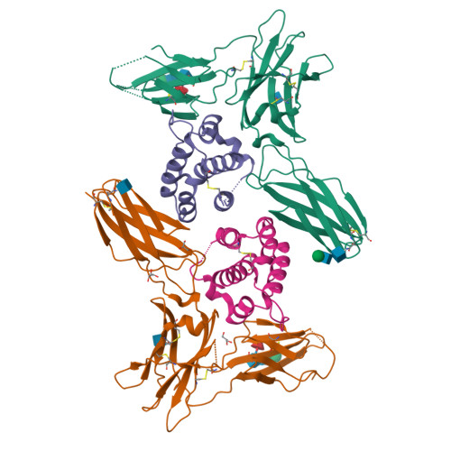
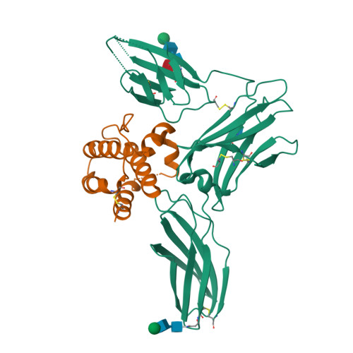
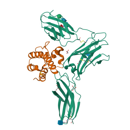
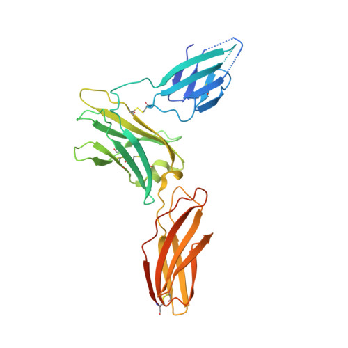
    5UV8, 5UWC

  • PubMed Abstract: 

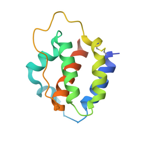
    The interleukin-3 (IL-3) receptor is a cell-surface heterodimer that links the haemopoietic, vascular and immune systems and is overexpressed in acute and chronic myeloid leukaemia progenitor cells. It belongs to the type I cytokine receptor family in which the α-subunits consist of two fibronectin III-like domains that bind cytokine, and a third, evolutionarily unrelated and topologically conserved, N-terminal domain (NTD) with unknown function. Here we show by crystallography that, while the NTD of IL3Rα is highly mobile in the presence of IL-3, it becomes surprisingly rigid in the presence of IL-3 K116W. Mutagenesis, biochemical and functional studies show that the NTD of IL3Rα regulates IL-3 binding and signalling and reveal an unexpected role in preventing spontaneous receptor dimerisation. Our work identifies a dual role for the NTD in this cytokine receptor family, protecting against inappropriate signalling and dynamically regulating cytokine receptor binding and function.


  • Organizational Affiliation

    Australian Cancer Research Foundation Rational Drug Discovery Centre, St. Vincent's Institute of Medical Research, Fitzroy, VIC, 3065, Australia.


Macromolecules
Find similar proteins by:  (by identity cutoff)  |  3D Structure
Entity ID: 1
MoleculeChains Sequence LengthOrganismDetailsImage
Interleukin-3 receptor subunit alphaA,
C [auth G]
288Homo sapiensMutation(s): 2 
Gene Names: IL3RAIL3R
UniProt & NIH Common Fund Data Resources
Find proteins for P26951 (Homo sapiens)
Explore P26951 
Go to UniProtKB:  P26951
PHAROS:  P26951
GTEx:  ENSG00000185291 
Entity Groups  
Sequence Clusters30% Identity50% Identity70% Identity90% Identity95% Identity100% Identity
UniProt GroupP26951
Glycosylation
Glycosylation Sites: 3Go to GlyGen: P26951-1
Sequence Annotations
Expand
  • Reference Sequence
Find similar proteins by:  (by identity cutoff)  |  3D Structure
Entity ID: 2
MoleculeChains Sequence LengthOrganismDetailsImage
Interleukin-3B,
D [auth I]
122Homo sapiensMutation(s): 1 
Gene Names: IL3
UniProt & NIH Common Fund Data Resources
Find proteins for P08700 (Homo sapiens)
Explore P08700 
Go to UniProtKB:  P08700
PHAROS:  P08700
GTEx:  ENSG00000164399 
Entity Groups  
Sequence Clusters30% Identity50% Identity70% Identity90% Identity95% Identity100% Identity
UniProt GroupP08700
Sequence Annotations
Expand
  • Reference Sequence
Oligosaccharides

Help

Entity ID: 3
MoleculeChains Length2D Diagram Glycosylation3D Interactions
beta-D-mannopyranose-(1-4)-2-acetamido-2-deoxy-beta-D-glucopyranose-(1-4)-2-acetamido-2-deoxy-beta-D-glucopyranoseE [auth C]3N-Glycosylation
Glycosylation Resources
GlyTouCan:  G15407YE
GlyCosmos:  G15407YE
GlyGen:  G15407YE
Entity ID: 4
MoleculeChains Length2D Diagram Glycosylation3D Interactions
beta-D-mannopyranose-(1-4)-2-acetamido-2-deoxy-beta-D-glucopyranose-(1-4)-[alpha-L-fucopyranose-(1-6)]2-acetamido-2-deoxy-beta-D-glucopyranoseF [auth D]4N-Glycosylation
Glycosylation Resources
GlyTouCan:  G32152BH
GlyCosmos:  G32152BH
GlyGen:  G32152BH
Entity ID: 5
MoleculeChains Length2D Diagram Glycosylation3D Interactions
beta-D-mannopyranose-(1-3)-2-acetamido-2-deoxy-beta-D-glucopyranose-(1-4)-[alpha-L-fucopyranose-(1-6)]2-acetamido-2-deoxy-beta-D-glucopyranoseG [auth E]4N-Glycosylation
Glycosylation Resources
GlyTouCan:  G27425HP
GlyCosmos:  G27425HP
GlyGen:  G27425HP
Experimental Data & Validation

Experimental Data

  • Method: X-RAY DIFFRACTION
  • Resolution: 2.70 Å
  • R-Value Free:  0.286 (Depositor), 0.290 (DCC) 
  • R-Value Work:  0.242 (Depositor), 0.240 (DCC) 
  • R-Value Observed: 0.244 (Depositor) 
Space Group: P 65 2 2
Unit Cell:
Length ( Å )Angle ( ˚ )
a = 132.039α = 90
b = 132.039β = 90
c = 210.631γ = 120
Software Package:
Software NamePurpose
REFMACrefinement
Aimlessdata scaling
XDSdata reduction
PHASERphasing

Structure Validation

View Full Validation Report


Ligand Structure Quality Assessment 

Created with Raphaël 2.3.0Worse 01 BetterLigand structure goodness of fit to experimental dataBest fitted NAGClick on this verticalbar to view details

Entry History 

Deposition Data

Revision History  (Full details and data files)

  • Version 1.0: 2018-02-07
    Type: Initial release
  • Version 2.0: 2020-07-29
    Type: Remediation
    Reason: Carbohydrate remediation
    Changes: Atomic model, Data collection, Derived calculations, Structure summary
  • Version 2.1: 2023-10-04
    Changes: Data collection, Database references, Refinement description, Structure summary
  • Version 2.2: 2024-11-13
    Changes: Structure summary