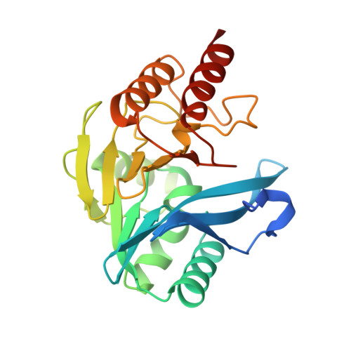
5A87

Crystal structure of the metallo-beta-lactamase VIM-5


Experimental Data Snapshot

  • Method: X-RAY DIFFRACTION
  • Resolution: 1.50 Å
  • R-Value Free: 0.192 
  • R-Value Work: 0.154 
  • R-Value Observed: 0.155 

Starting Model: experimental
View more details

wwPDB Validation   3D Report Full Report


This is version 1.2 of the entry. See complete history


Literature

Comparison of Verona Integron-Borne Metallo-beta-Lactamase (VIM) Variants Reveals Differences in Stability and Inhibition Profiles.

Makena, A.Duzgun, A.O.Brem, J.McDonough, M.A.Rydzik, A.M.Abboud, M.I.Saral, A.Cicek, A.C.Sandalli, C.Schofield, C.J.

(2015) Antimicrob Agents Chemother 60: 1377-1384

  • DOI: https://doi.org/10.1128/AAC.01768-15
  • Primary Citation of Related Structures:  
    5A87

  • PubMed Abstract: 

    Metallo-β-lactamases (MBLs) are of increasing clinical significance; the development of clinically useful MBL inhibitors is challenged by the rapid evolution of variant MBLs. The Verona integron-borne metallo-β-lactamase (VIM) enzymes are among the most widely distributed MBLs, with >40 VIM variants having been reported. We report on the crystallographic analysis of VIM-5 and comparison of biochemical and biophysical properties of VIM-1, VIM-2, VIM-4, VIM-5, and VIM-38. Recombinant VIM variants were produced and purified, and their secondary structure and thermal stabilities were investigated by circular dichroism analyses. Steady-state kinetic analyses with a representative panel of β-lactam substrates were carried out to compare the catalytic efficiencies of the VIM variants. Furthermore, a set of metalloenzyme inhibitors were screened to compare their effects on the different VIM variants. The results reveal only small variations in the kinetic parameters of the VIM variants but substantial differences in their thermal stabilities and inhibition profiles. Overall, these results support the proposal that protein stability may be a factor in MBL evolution and highlight the importance of screening MBL variants during inhibitor development programs.


  • Organizational Affiliation

    Chemistry Research Laboratory, Department of Chemistry, University of Oxford, Oxford, United Kingdom.


Macromolecules
Find similar proteins by:  (by identity cutoff)  |  3D Structure
Entity ID: 1
MoleculeChains Sequence LengthOrganismDetailsImage
METALLO-BETA-LACTAMASE VIM-5
A, B
248Klebsiella pneumoniaeMutation(s): 0 
EC: 3.5.2.6
UniProt
Find proteins for Q8GKX2 (Klebsiella pneumoniae)
Explore Q8GKX2 
Go to UniProtKB:  Q8GKX2
Entity Groups  
Sequence Clusters30% Identity50% Identity70% Identity90% Identity95% Identity100% Identity
UniProt GroupQ8GKX2
Sequence Annotations
Expand
  • Reference Sequence
Small Molecules
Ligands 2 Unique
IDChains Name / Formula / InChI Key2D Diagram3D Interactions
GOL
Query on GOL

Download Ideal Coordinates CCD File 
C [auth A],
E [auth A],
G [auth A],
J [auth B]
GLYCEROL
C3 H8 O3
PEDCQBHIVMGVHV-UHFFFAOYSA-N
ZN
Query on ZN

Download Ideal Coordinates CCD File 
D [auth A],
F [auth A],
H [auth A],
I [auth B],
K [auth B]
ZINC ION
Zn
PTFCDOFLOPIGGS-UHFFFAOYSA-N
Experimental Data & Validation

Experimental Data

  • Method: X-RAY DIFFRACTION
  • Resolution: 1.50 Å
  • R-Value Free: 0.192 
  • R-Value Work: 0.154 
  • R-Value Observed: 0.155 
  • Space Group: P 1 21 1
Unit Cell:
Length ( Å )Angle ( ˚ )
a = 41.27α = 90
b = 78.96β = 104.82
c = 64.03γ = 90
Software Package:
Software NamePurpose
PHENIXrefinement
MOSFLMdata reduction
SCALEPACKdata scaling
PHASERphasing

Structure Validation

View Full Validation Report


Entry History 

Deposition Data

Revision History  (Full details and data files)

  • Version 1.0: 2015-12-23
    Type: Initial release
  • Version 1.1: 2017-03-22
    Changes: Database references
  • Version 1.2: 2024-01-10
    Changes: Data collection, Database references, Derived calculations, Other, Refinement description