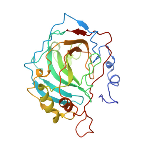
5CAC

REFINED STRUCTURE OF HUMAN CARBONIC ANHYDRASE II AT 2.0 ANGSTROMS RESOLUTION


Experimental Data Snapshot

  • Method: X-RAY DIFFRACTION
  • Resolution: 2.20 Å
  • R-Value Observed: 0.189 

wwPDB Validation   3D Report Full Report


This is version 1.4 of the entry. See complete history


Literature

Refined structure of human carbonic anhydrase II at 2.0 A resolution.

Eriksson, A.E.Jones, T.A.Liljas, A.

(1988) Proteins 4: 274-282

  • DOI: https://doi.org/10.1002/prot.340040406
  • Primary Citation of Related Structures:  
    1CA2, 4CAC, 5CAC

  • PubMed Abstract: 

    The structure of human erythrocytic carbonic anhydrase II has been refined by constrained and restrained structure-factor least-squares refinement at 2.0 A resolution. The conventional crystallographic R value is 17.3%. Of 167 solvent molecules associated with the protein, four are buried and stabilize secondary structure elements. The zinc ion is ligated to three histidyl residues and one water molecule in a nearly tetrahedral geometry. In addition to the zinc-bound water, seven more water molecules are identified in the active site. Assuming that Glu-106 is deprotonated at pH 8.5, some of the hydrogen bond donor-acceptor relations in the active site can be assigned and are described here in detail. The O gamma 1 atom of Thr-199 donates its proton to the O epsilon 1 atom of Glu-106 and can function as a hydrogen bond acceptor only in additional hydrogen bonds.


  • Organizational Affiliation

    Department of Molecular Biology, Biomedical Center, Uppsala, Sweden.


Macromolecules
Find similar proteins by:  (by identity cutoff)  |  3D Structure
Entity ID: 1
MoleculeChains Sequence LengthOrganismDetailsImage
CARBONIC ANHYDRASE FORM C259Homo sapiensMutation(s): 0 
EC: 4.2.1.1 (PDB Primary Data), 4.2.1.69 (UniProt)
UniProt & NIH Common Fund Data Resources
Find proteins for P00918 (Homo sapiens)
Explore P00918 
Go to UniProtKB:  P00918
PHAROS:  P00918
GTEx:  ENSG00000104267 
Entity Groups  
Sequence Clusters30% Identity50% Identity70% Identity90% Identity95% Identity100% Identity
UniProt GroupP00918
Sequence Annotations
Expand
  • Reference Sequence
Small Molecules
Ligands 2 Unique
IDChains Name / Formula / InChI Key2D Diagram3D Interactions
SO3
Query on SO3

Download Ideal Coordinates CCD File 
C [auth A]SULFITE ION
O3 S
LSNNMFCWUKXFEE-UHFFFAOYSA-L
ZN
Query on ZN

Download Ideal Coordinates CCD File 
B [auth A]ZINC ION
Zn
PTFCDOFLOPIGGS-UHFFFAOYSA-N
Experimental Data & Validation

Experimental Data

  • Method: X-RAY DIFFRACTION
  • Resolution: 2.20 Å
  • R-Value Observed: 0.189 
  • Space Group: P 1 21 1
Unit Cell:
Length ( Å )Angle ( ˚ )
a = 42.7α = 90
b = 41.7β = 104.6
c = 73γ = 90
Software Package:
Software NamePurpose
PROLSQrefinement

Structure Validation

View Full Validation Report


Entry History 

Deposition Data

Revision History  (Full details and data files)

  • Version 1.0: 1994-01-31
    Type: Initial release
  • Version 1.1: 2008-03-03
    Changes: Version format compliance
  • Version 1.2: 2011-07-13
    Changes: Version format compliance
  • Version 1.3: 2017-11-29
    Changes: Derived calculations, Other
  • Version 1.4: 2024-03-06
    Changes: Data collection, Database references, Derived calculations