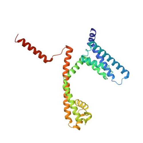
5HVX

Full length Wild-Type Open-form Sodium Channel NavMs


Experimental Data Snapshot

  • Method: X-RAY DIFFRACTION
  • Resolution: 2.45 Å
  • R-Value Free: 0.233 
  • R-Value Work: 0.207 
  • R-Value Observed: 0.209 

Starting Model: experimental
View more details

wwPDB Validation   3D Report Full Report


Ligand Structure Quality Assessment 


This is version 1.2 of the entry. See complete history


Literature

The complete structure of an activated open sodium channel.

Sula, A.Booker, J.Ng, L.C.Naylor, C.E.DeCaen, P.G.Wallace, B.A.

(2017) Nat Commun 8: 14205-14205

  • DOI: https://doi.org/10.1038/ncomms14205
  • Primary Citation of Related Structures:  
    5HVD, 5HVX

  • PubMed Abstract: 

    Voltage-gated sodium channels (Navs) play essential roles in excitable tissues, with their activation and opening resulting in the initial phase of the action potential. The cycling of Navs through open, closed and inactivated states, and their closely choreographed relationships with the activities of other ion channels lead to exquisite control of intracellular ion concentrations in both prokaryotes and eukaryotes. Here we present the 2.45 Å resolution crystal structure of the complete NavMs prokaryotic sodium channel in a fully open conformation. A canonical activated conformation of the voltage sensor S4 helix, an open selectivity filter leading to an open activation gate at the intracellular membrane surface and the intracellular C-terminal domain are visible in the structure. It includes a heretofore unseen interaction motif between W77 of S3, the S4-S5 interdomain linker, and the C-terminus, which is associated with regulation of opening and closing of the intracellular gate.


  • Organizational Affiliation

    Institute of Structural and Molecular Biology, Birkbeck College, University of London, Malet Street, London WC1E 7HX, UK.


Macromolecules
Find similar proteins by:  (by identity cutoff)  |  3D Structure
Entity ID: 1
MoleculeChains Sequence LengthOrganismDetailsImage
Ion transport protein277Magnetococcus marinus MC-1Mutation(s): 0 
Gene Names: Mmc1_0798
Membrane Entity: Yes 
UniProt
Find proteins for A0L5S6 (Magnetococcus marinus (strain ATCC BAA-1437 / JCM 17883 / MC-1))
Explore A0L5S6 
Go to UniProtKB:  A0L5S6
Entity Groups  
Sequence Clusters30% Identity50% Identity70% Identity90% Identity95% Identity100% Identity
UniProt GroupA0L5S6
Sequence Annotations
Expand
  • Reference Sequence
Small Molecules
Ligands 5 Unique
IDChains Name / Formula / InChI Key2D Diagram3D Interactions
12P
Query on 12P

Download Ideal Coordinates CCD File 
F [auth A]DODECAETHYLENE GLYCOL
C24 H50 O13
WRZXKWFJEFFURH-UHFFFAOYSA-N
2CV
Query on 2CV

Download Ideal Coordinates CCD File 
E [auth A]HEGA-10
C18 H37 N O7
ITEIKACYSCODFV-ATLSCFEFSA-N
MES
Query on MES

Download Ideal Coordinates CCD File 
H [auth A],
I [auth A]
2-(N-MORPHOLINO)-ETHANESULFONIC ACID
C6 H13 N O4 S
SXGZJKUKBWWHRA-UHFFFAOYSA-N
EDO
Query on EDO

Download Ideal Coordinates CCD File 
G [auth A]1,2-ETHANEDIOL
C2 H6 O2
LYCAIKOWRPUZTN-UHFFFAOYSA-N
NA
Query on NA

Download Ideal Coordinates CCD File 
B [auth A],
C [auth A],
D [auth A]
SODIUM ION
Na
FKNQFGJONOIPTF-UHFFFAOYSA-N
Experimental Data & Validation

Experimental Data

  • Method: X-RAY DIFFRACTION
  • Resolution: 2.45 Å
  • R-Value Free: 0.233 
  • R-Value Work: 0.207 
  • R-Value Observed: 0.209 
  • Space Group: I 4 2 2
Unit Cell:
Length ( Å )Angle ( ˚ )
a = 109α = 90
b = 109β = 90
c = 210.59γ = 90
Software Package:
Software NamePurpose
BUSTERrefinement
XDSdata reduction
XDSdata scaling
PHASERphasing

Structure Validation

View Full Validation Report


Ligand Structure Quality Assessment 


Entry History & Funding Information

Deposition Data


Funding OrganizationLocationGrant Number
Biotechnology and Biological Sciences Research CouncilUnited KingdomBB/L02625/21
Biotechnology and Biological Sciences Research CouncilUnited KingdomBB/L006790

Revision History  (Full details and data files)

  • Version 1.0: 2017-03-01
    Type: Initial release
  • Version 1.1: 2017-08-30
    Changes: Author supporting evidence
  • Version 1.2: 2024-01-10
    Changes: Data collection, Database references, Refinement description