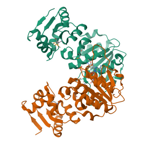
Crystal Structure of AdoMet bound rRNA methyltransferase from Sinorhizobium meliloti
Dey, D., Hegde, R.P., Almo, S.C., Ramakumar, S., Ramagopal, U.A.To be published.
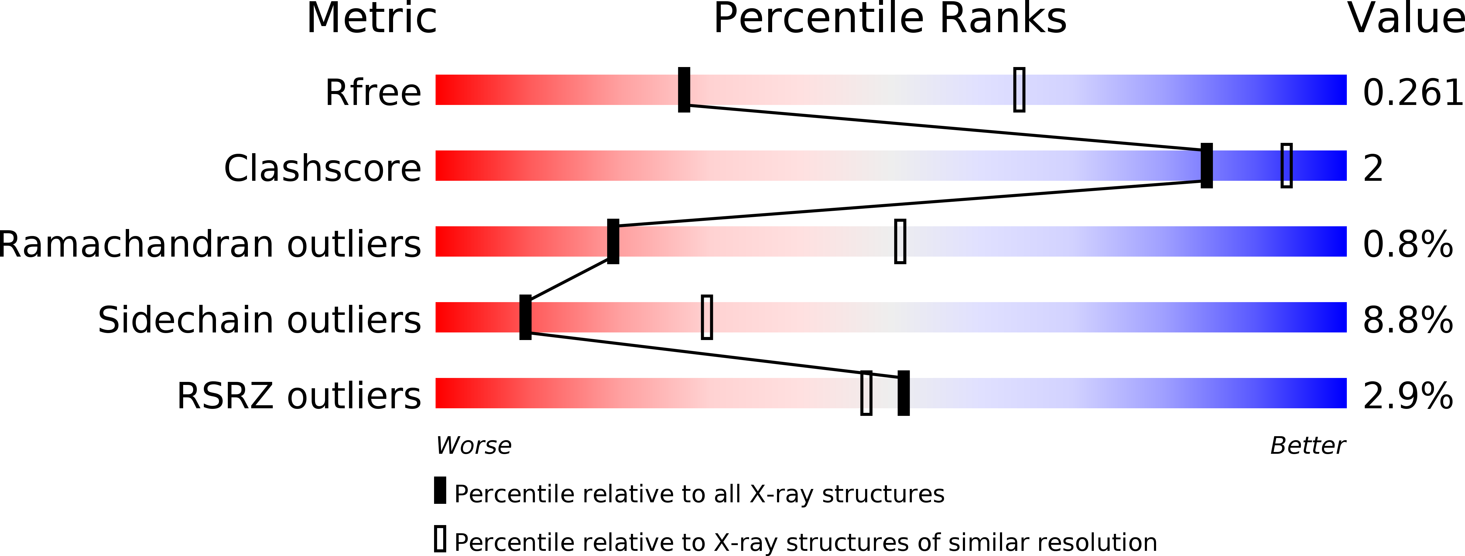
Experimental Data Snapshot
Entity ID: 1 | |||||
|---|---|---|---|---|---|
| Molecule | Chains | Sequence Length | Organism | Details | Image |
| Probable RNA methyltransferase, TrmH family | 288 | Sinorhizobium meliloti 1021 | Mutation(s): 0 Gene Names: SMc01130 EC: 2.1.1 | 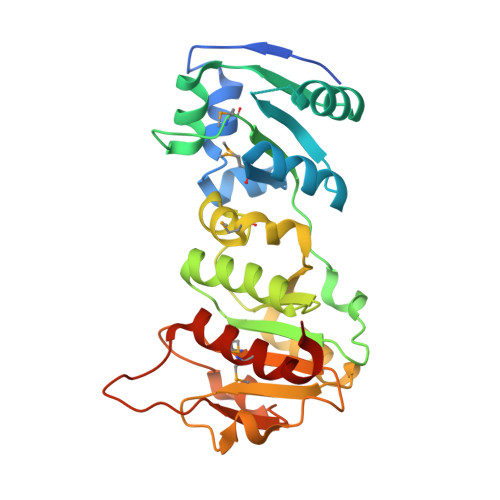 | |
UniProt | |||||
Find proteins for Q92SJ4 (Rhizobium meliloti (strain 1021)) Explore Q92SJ4 Go to UniProtKB: Q92SJ4 | |||||
Entity Groups | |||||
| Sequence Clusters | 30% Identity50% Identity70% Identity90% Identity95% Identity100% Identity | ||||
| UniProt Group | Q92SJ4 | ||||
Sequence AnnotationsExpand | |||||
| |||||
| Ligands 3 Unique | |||||
|---|---|---|---|---|---|
| ID | Chains | Name / Formula / InChI Key | 2D Diagram | 3D Interactions | |
| SAM Query on SAM | F [auth A], J [auth B] | S-ADENOSYLMETHIONINE C15 H22 N6 O5 S MEFKEPWMEQBLKI-FCKMPRQPSA-N |  | ||
| SO4 Query on SO4 | E [auth A], I [auth B] | SULFATE ION O4 S QAOWNCQODCNURD-UHFFFAOYSA-L |  | ||
| CO Query on CO | C [auth A], D [auth A], G [auth B], H [auth B] | COBALT (II) ION Co XLJKHNWPARRRJB-UHFFFAOYSA-N |  | ||
| Modified Residues 1 Unique | |||||
|---|---|---|---|---|---|
| ID | Chains | Type | Formula | 2D Diagram | Parent |
| MSE Query on MSE | A, B | L-PEPTIDE LINKING | C5 H11 N O2 Se |  | MET |
| Length ( Å ) | Angle ( ˚ ) |
|---|---|
| a = 64.855 | α = 90 |
| b = 82.896 | β = 90 |
| c = 108.806 | γ = 90 |
| Software Name | Purpose |
|---|---|
| HKL-3000 | data processing |
| HKL-3000 | data scaling |
| REFMAC | refinement |
RCSB PDB is hosted by

RCSB PDB is a member of the