5NJV

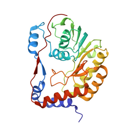
Flavivirus NS5 domain


Experimental Data Snapshot

  • Method: X-RAY DIFFRACTION
  • Resolution: 2.00 Å
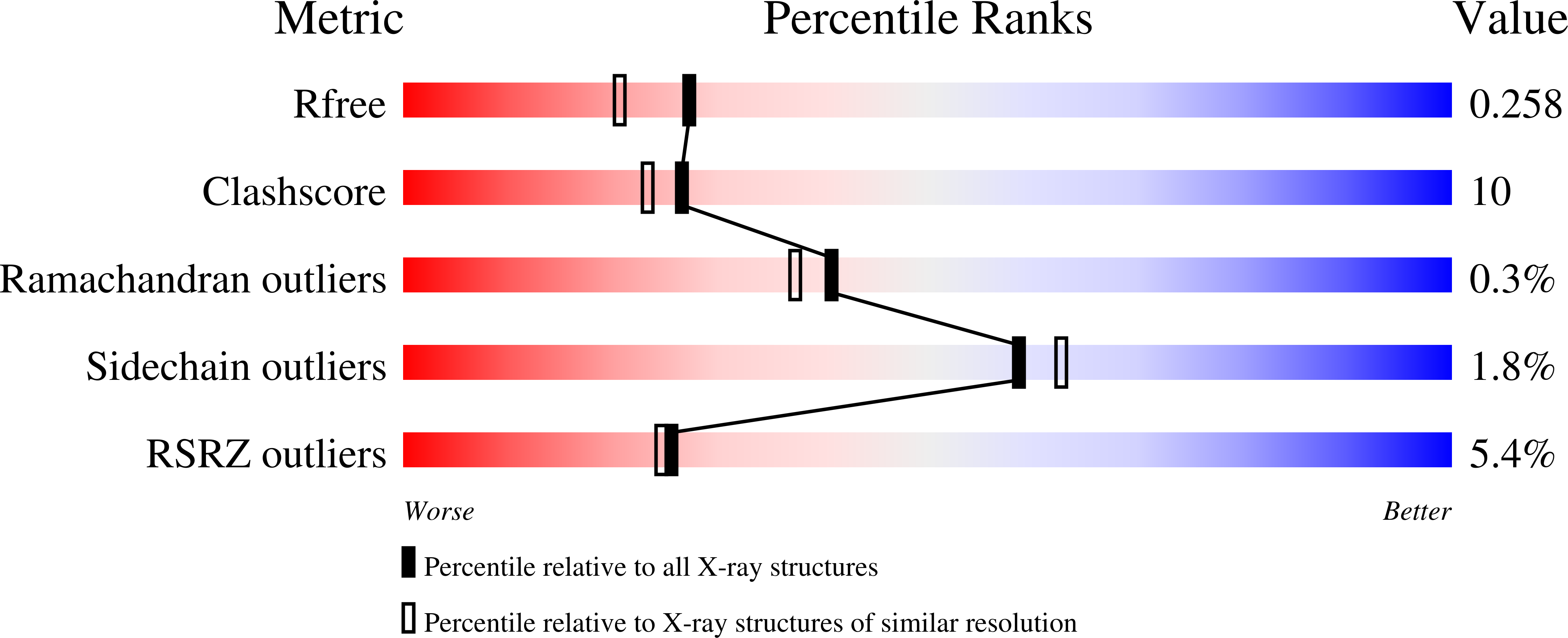
  • R-Value Free: 0.258 
  • R-Value Work: 0.201 
  • R-Value Observed: 0.204 

Starting Model: experimental
View more details

wwPDB Validation   3D Report Full Report


Ligand Structure Quality Assessment 


This is version 1.3 of the entry. See complete history


Literature

The structure of the binary methyltransferase-SAH complex from Zika virus reveals a novel conformation for the mechanism of mRNA capping.

Chatrin, C.Talapatra, S.K.Canard, B.Kozielski, F.

(2018) Oncotarget 9: 3160-3171

  • DOI: https://doi.org/10.18632/oncotarget.23223
  • Primary Citation of Related Structures:  
    5NJU, 5NJV

  • PubMed Abstract: 

    Zika virus, a flavivirus like Dengue and West Nile viruses, poses a significant risk as a pathogen in the category of emerging infectious diseases. Zika infections typically cause nonspecific, mild symptoms, but can also manifest as a neurological disorder like Guillain-Barré syndrome. Infection in pregnant women is linked to microcephaly in newborn infants. The methyltransferase domain of the non-structural protein 5 is responsible for two sequential methylations of the 5'-RNA cap. This is crucial for genome stability, efficient translation, and escape from the host immune response. Here we present the crystal structures of the Zika methyltransferase domain in complex with the methyl-donor SAM and its by-product SAH. The methyltransferase-SAH binary complex presents a new conformation of a "closed" or "obstructed" state that would restrict the binding of new RNA for capping. The combination and comparison of our new structures with recently published Zika methyltransferase structures provide a first glimpse into the structural mechanism of Zika virus mRNA capping.


  • Organizational Affiliation

    Department of Pharmaceutical and Biological Chemistry, UCL School of Pharmacy, WC1N 1AX, London, United Kingdom.


Macromolecules
Find similar proteins by:  (by identity cutoff)  |  3D Structure
Entity ID: 1
MoleculeChains Sequence LengthOrganismDetailsImage
NS5
A, B, C, D
262Zika virusMutation(s): 0 
UniProt
Find proteins for A0A024B7W1 (Zika virus (isolate ZIKV/Human/French Polynesia/10087PF/2013))
Explore A0A024B7W1 
Go to UniProtKB:  A0A024B7W1
Entity Groups  
Sequence Clusters30% Identity50% Identity70% Identity90% Identity95% Identity100% Identity
UniProt GroupA0A024B7W1
Sequence Annotations
Expand
  • Reference Sequence
Small Molecules
Ligands 2 Unique
IDChains Name / Formula / InChI Key2D Diagram3D Interactions
SAM
Query on SAM

Download Ideal Coordinates CCD File 
E [auth A],
H [auth B],
K [auth C],
L [auth D]
S-ADENOSYLMETHIONINE
C15 H22 N6 O5 S
MEFKEPWMEQBLKI-FCKMPRQPSA-N
CL
Query on CL

Download Ideal Coordinates CCD File 
F [auth A],
G [auth A],
I [auth B],
J [auth B]
CHLORIDE ION
Cl
VEXZGXHMUGYJMC-UHFFFAOYSA-M
Experimental Data & Validation

Experimental Data

  • Method: X-RAY DIFFRACTION
  • Resolution: 2.00 Å
  • R-Value Free: 0.258 
  • R-Value Work: 0.201 
  • R-Value Observed: 0.204 
  • Space Group: P 61
Unit Cell:
Length ( Å )Angle ( ˚ )
a = 125.92α = 90
b = 125.92β = 90
c = 125.854γ = 120
Software Package:
Software NamePurpose
PHENIXrefinement
iMOSFLMdata reduction
SCALAdata scaling
PHASERphasing

Structure Validation

View Full Validation Report


Ligand Structure Quality Assessment 


Entry History 

Deposition Data

Revision History  (Full details and data files)

  • Version 1.0: 2018-01-24
    Type: Initial release
  • Version 1.1: 2018-04-18
    Changes: Data collection, Database references
  • Version 1.2: 2024-01-17
    Changes: Data collection, Database references, Derived calculations, Refinement description
  • Version 1.3: 2024-11-20
    Changes: Structure summary