CHARACTERISATION OF A FURANOSE SPECIFIC ABC TRANSPORTER ESSENTIAL FOR ARABINOSE UTILISATION FROM THE LIGNOCELLULOSE DEGRADING BACTERIUM SHEWANELLA SP. ANA-3
Herman, R., Drousiotis, K., Wilkinson, A.J., Thomas, G.H.To be published.
Experimental Data Snapshot
Starting Model: experimental
View more details
Entity ID: 1 | |||||
|---|---|---|---|---|---|
| Molecule | Chains | Sequence Length | Organism | Details | Image |
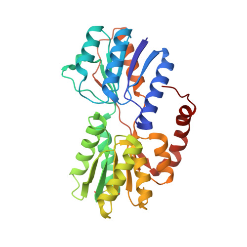
| Periplasmic binding protein/LacI transcriptional regulator | 302 | Shewanella sp. ANA-3 | Mutation(s): 0 Gene Names: Shewana3_2073 |  | |
UniProt | |||||
Find proteins for A0KWY4 (Shewanella sp. (strain ANA-3)) Explore A0KWY4 Go to UniProtKB: A0KWY4 | |||||
Entity Groups | |||||
| Sequence Clusters | 30% Identity50% Identity70% Identity90% Identity95% Identity100% Identity | ||||
| UniProt Group | A0KWY4 | ||||
Sequence AnnotationsExpand | |||||
| |||||
| Ligands 4 Unique | |||||
|---|---|---|---|---|---|
| ID | Chains | Name / Formula / InChI Key | 2D Diagram | 3D Interactions | |
| FUB (Subject of Investigation/LOI) Query on FUB | J [auth A], O [auth B] | beta-L-arabinofuranose C5 H10 O5 HMFHBZSHGGEWLO-KLVWXMOXSA-N |  | ||
| AHR (Subject of Investigation/LOI) Query on AHR | I [auth A], N [auth B] | alpha-L-arabinofuranose C5 H10 O5 HMFHBZSHGGEWLO-QMKXCQHVSA-N |  | ||
| GOL Query on GOL | C [auth A] D [auth A] E [auth A] F [auth A] G [auth A] | GLYCEROL C3 H8 O3 PEDCQBHIVMGVHV-UHFFFAOYSA-N |  | ||
| ACT Query on ACT | M [auth B] | ACETATE ION C2 H3 O2 QTBSBXVTEAMEQO-UHFFFAOYSA-M |  | ||
| Length ( Å ) | Angle ( ˚ ) |
|---|---|
| a = 73.917 | α = 90 |
| b = 86.327 | β = 90 |
| c = 87.282 | γ = 90 |
| Software Name | Purpose |
|---|---|
| REFMAC | refinement |
| Aimless | data scaling |
| PDB_EXTRACT | data extraction |
| MOLREP | phasing |
| Funding Organization | Location | Grant Number |
|---|---|---|
| Biotechnology and Biological Sciences Research Council | United Kingdom | BB/N01040X/1 |