5OK4

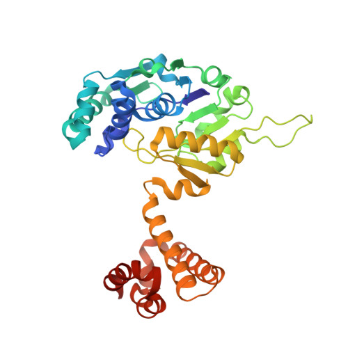
Crystal structure of native [Fe]-hydrogenase Hmd from Methanothermobacter marburgensis inactivated by O2.


Experimental Data Snapshot

  • Method: X-RAY DIFFRACTION
  • Resolution: 1.29 Å
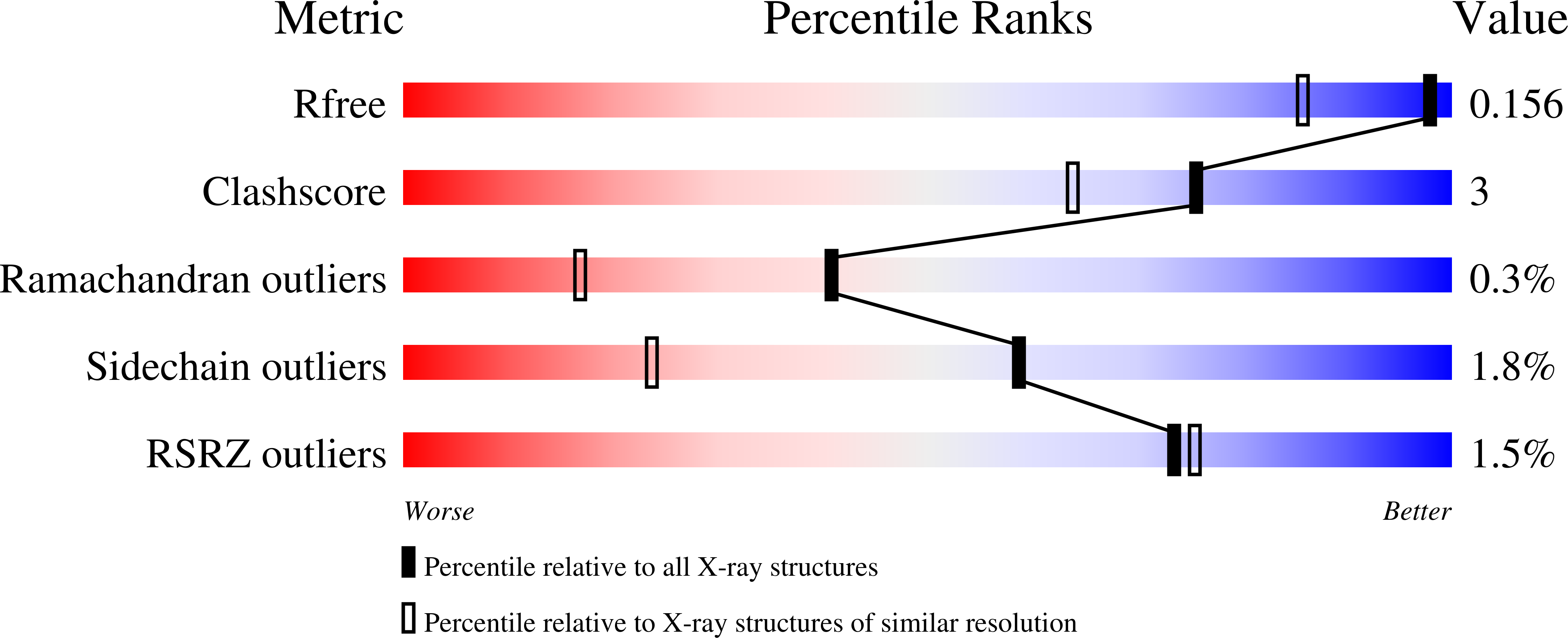
  • R-Value Free: 0.147 
  • R-Value Work: 0.127 
  • R-Value Observed: 0.128 

Starting Model: experimental
View more details

wwPDB Validation   3D Report Full Report


Ligand Structure Quality Assessment 


This is version 1.2 of the entry. See complete history


Literature

Dioxygen Sensitivity of [Fe]-Hydrogenase in the Presence of Reducing Substrates.

Huang, G.Wagner, T.Ermler, U.Bill, E.Ataka, K.Shima, S.

(2018) Angew Chem Int Ed Engl 57: 4917-4920

  • DOI: https://doi.org/10.1002/anie.201712293
  • Primary Citation of Related Structures:  
    5OK4

  • PubMed Abstract: 

    Mono-iron hydrogenase ([Fe]-hydrogenase) reversibly catalyzes the transfer of a hydride ion from H 2 to methenyltetrahydromethanopterin (methenyl-H 4 MPT + ) to form methylene-H 4 MPT. Its iron guanylylpyridinol (FeGP) cofactor plays a key role in H 2 activation. Evidence is presented for O 2 sensitivity of [Fe]-hydrogenase under turnover conditions in the presence of reducing substrates, methylene-H 4 MPT or methenyl-H 4 MPT + /H 2 . Only then, H 2 O 2 is generated, which decomposes the FeGP cofactor; as demonstrated by spectroscopic analyses and the crystal structure of the deactivated enzyme. O 2 reduction to H 2 O 2 requires a reductant, which can be a catalytic intermediate transiently formed during the [Fe]-hydrogenase reaction. The most probable candidate is an iron hydride species; its presence has already been predicted by theoretical studies of the catalytic reaction. The findings support predictions because the same type of reduction reaction is described for ruthenium hydride complexes that hydrogenate polar compounds.


  • Organizational Affiliation

    Max-Planck-Institut für terrestrische Mikrobiologie, Karl-von-Frisch-Straße 10, 35043, Marburg, Germany.


Macromolecules
Find similar proteins by:  (by identity cutoff)  |  3D Structure
Entity ID: 1
MoleculeChains Sequence LengthOrganismDetailsImage
5,10-methenyltetrahydromethanopterin hydrogenase344Methanothermobacter marburgensis str. MarburgMutation(s): 0 
EC: 1.12.98.2
UniProt
Find proteins for P32440 (Methanothermobacter marburgensis (strain ATCC BAA-927 / DSM 2133 / JCM 14651 / NBRC 100331 / OCM 82 / Marburg))
Explore P32440 
Go to UniProtKB:  P32440
Entity Groups  
Sequence Clusters30% Identity50% Identity70% Identity90% Identity95% Identity100% Identity
UniProt GroupP32440
Sequence Annotations
Expand
  • Reference Sequence
Experimental Data & Validation

Experimental Data

  • Method: X-RAY DIFFRACTION
  • Resolution: 1.29 Å
  • R-Value Free: 0.147 
  • R-Value Work: 0.127 
  • R-Value Observed: 0.128 
  • Space Group: H 3 2
Unit Cell:
Length ( Å )Angle ( ˚ )
a = 127.418α = 90
b = 127.418β = 90
c = 141.211γ = 120
Software Package:
Software NamePurpose
PHENIXrefinement
XDSdata reduction
SCALAdata scaling
PHASERphasing

Structure Validation

View Full Validation Report


Ligand Structure Quality Assessment 


Entry History 

Deposition Data

Revision History  (Full details and data files)

  • Version 1.0: 2018-05-16
    Type: Initial release
  • Version 1.1: 2018-05-30
    Changes: Data collection, Database references
  • Version 1.2: 2024-01-17
    Changes: Data collection, Database references, Derived calculations, Refinement description