5OWP

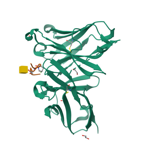
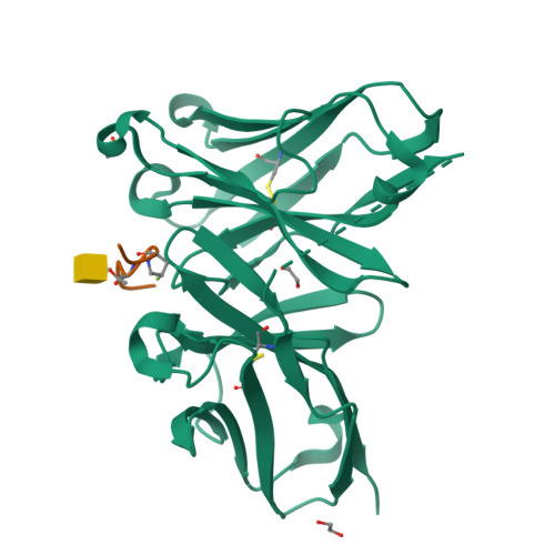
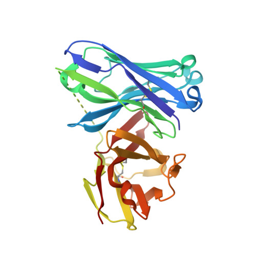
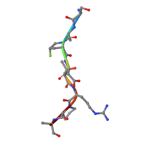
Crystal structure of glycopeptide "GVTSAfPDT*RPAP" in complex with scFv-SM3


Experimental Data Snapshot

  • Method: X-RAY DIFFRACTION
  • Resolution: 1.85 Å
  • R-Value Free: 0.256 
  • R-Value Work: 0.194 
  • R-Value Observed: 0.196 

Starting Model: experimental
View more details

wwPDB Validation   3D Report Full Report


Ligand Structure Quality Assessment 


This is version 2.2 of the entry. See complete history


Literature

The Use of Fluoroproline in MUC1 Antigen Enables Efficient Detection of Antibodies in Patients with Prostate Cancer.

Somovilla, V.J.Bermejo, I.A.Albuquerque, I.S.Martinez-Saez, N.Castro-Lopez, J.Garcia-Martin, F.Companon, I.Hinou, H.Nishimura, S.I.Jimenez-Barbero, J.Asensio, J.L.Avenoza, A.Busto, J.H.Hurtado-Guerrero, R.Peregrina, J.M.Bernardes, G.J.L.Corzana, F.

(2017) J Am Chem Soc 139: 18255-18261

  • DOI: https://doi.org/10.1021/jacs.7b09447
  • Primary Citation of Related Structures:  
    5OWP

  • PubMed Abstract: 

    A structure-based design of a new generation of tumor-associated glycopeptides with improved affinity against two anti-MUC1 antibodies is described. These unique antigens feature a fluorinated proline residue, such as a (4S)-4-fluoro-l-proline or 4,4-difluoro-l-proline, at the most immunogenic domain. Binding assays using biolayer interferometry reveal 3-fold to 10-fold affinity improvement with respect to the natural (glyco)peptides. According to X-ray crystallography and MD simulations, the fluorinated residues stabilize the antigen-antibody complex by enhancing key CH/π interactions. Interestingly, a notable improvement in detection of cancer-associated anti-MUC1 antibodies from serum of patients with prostate cancer is achieved with the non-natural antigens, which proves that these derivatives can be considered better diagnostic tools than the natural antigen for prostate cancer.


  • Organizational Affiliation

    Departamento de Química, Universidad de La Rioja, Centro de Investigación en Síntesis Química , 26006 Logroño, Spain.


Macromolecules
Find similar proteins by:  (by identity cutoff)  |  3D Structure
Entity ID: 1
MoleculeChains Sequence LengthOrganismDetailsImage
Ig heavy chain V-III region J606,Ig lambda-1 chain V region H2020A [auth H]244Mus musculusMutation(s): 0 
UniProt
Find proteins for P01726 (Mus musculus)
Explore P01726 
Go to UniProtKB:  P01726
Find proteins for P01801 (Mus musculus)
Explore P01801 
Go to UniProtKB:  P01801
Entity Groups  
Sequence Clusters30% Identity50% Identity70% Identity90% Identity95% Identity100% Identity
UniProt GroupsP01726P01801
Sequence Annotations
Expand
  • Reference Sequence

Find similar proteins by:  Sequence   |   3D Structure  

Entity ID: 2
MoleculeChains Sequence LengthOrganismDetailsImage
5,6-DIHYDRO-BENZO[H]CINNOLIN-3-YLAMINEB [auth D]8synthetic constructMutation(s): 0 
Glycosylation
Glycosylation Sites: 1
Sequence Annotations
Expand
  • Reference Sequence
Experimental Data & Validation

Experimental Data

  • Method: X-RAY DIFFRACTION
  • Resolution: 1.85 Å
  • R-Value Free: 0.256 
  • R-Value Work: 0.194 
  • R-Value Observed: 0.196 
  • Space Group: P 21 21 21
Unit Cell:
Length ( Å )Angle ( ˚ )
a = 35.399α = 90
b = 68.151β = 90
c = 90.358γ = 90
Software Package:
Software NamePurpose
REFMACrefinement
XDSdata reduction
SCALAdata scaling
PHASERphasing

Structure Validation

View Full Validation Report


Ligand Structure Quality Assessment 


Entry History 

Revision History  (Full details and data files)

  • Version 1.0: 2017-12-13
    Type: Initial release
  • Version 1.1: 2017-12-27
    Changes: Database references
  • Version 2.0: 2020-07-29
    Type: Remediation
    Reason: Carbohydrate remediation
    Changes: Atomic model, Data collection, Derived calculations, Structure summary
  • Version 2.1: 2024-01-17
    Changes: Data collection, Database references, Derived calculations, Refinement description, Structure summary
  • Version 2.2: 2024-10-23
    Changes: Structure summary