5T05

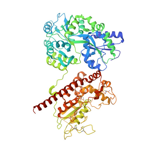
Crystal structure of heparan sulfate 6-O-sulfotransferase with bound PAP and IdoA2S containing hexasaccharide substrate


Experimental Data Snapshot

  • Method: X-RAY DIFFRACTION
  • Resolution: 1.95 Å
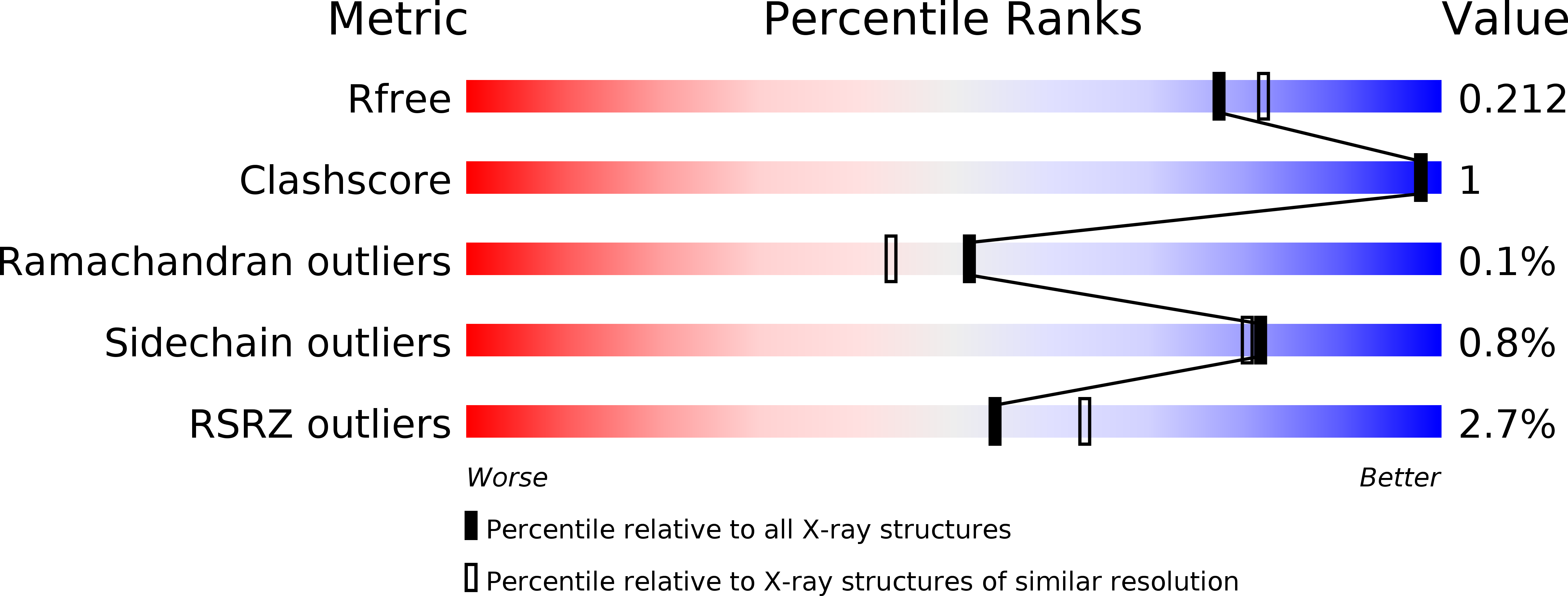
  • R-Value Free: 0.212 
  • R-Value Work: 0.178 
  • R-Value Observed: 0.179 

wwPDB Validation   3D Report Full Report


Ligand Structure Quality Assessment 


This is version 2.1 of the entry. See complete history


Literature

Structure Based Substrate Specificity Analysis of Heparan Sulfate 6-O-Sulfotransferases.

Xu, Y.Moon, A.F.Xu, S.Krahn, J.M.Liu, J.Pedersen, L.C.

(2017) ACS Chem Biol 12: 73-82

  • DOI: https://doi.org/10.1021/acschembio.6b00841
  • Primary Citation of Related Structures:  
    5T03, 5T05, 5T0A

  • PubMed Abstract: 

    Heparan sulfate (HS) is a sulfated polysaccharide exhibiting essential physiological functions. HS 6-O-sulfotransferase (6-OST) transfers a sulfo group to the 6-OH position of glucosamine units to confer a variety of HS biological activities. There are three different isoforms of 6-OST in the human genome. Here, we report crystal structures of the ternary complex of 6-OST with the sulfo donor analog 3'-phosphoadenosine 5'-phosphate and three different oligosaccharide substrates at 1.95 to 2.1 Å resolutions. Structural and mutational analyses reveal amino acid residues that contribute to catalysis and substrate recognition of 6-OST. Unexpectedly, the structures reveal 6-OST engages HS in a completely different orientation than other HS sulfotransferases and sheds light on the basic HS requirements for specificity. These findings also contribute structural information to understand mutations in human 6-OST isoform 1 associated with the human genetic disease idiopathic hypogonadotropic hypogonadism characterized by incomplete or lack of puberty.


  • Organizational Affiliation

    Division of Chemical Biology and Medicinal Chemistry, Eshelman School of Pharmacy, University of North Carolina , Chapel Hill, North Carolina 27599, United States.


Macromolecules
Find similar proteins by:  (by identity cutoff)  |  3D Structure
Entity ID: 1
MoleculeChains Sequence LengthOrganismDetailsImage
maltose binding protein - heparan sulfate 6-O-sulfotransferase isoform 3 fusion protein
A, B
692Escherichia coli O157:H7Danio rerio
This entity is chimeric
Mutation(s): 8 
Gene Names: malEZ5632ECs5017hs6st3
EC: 2.8.2
UniProt
Find proteins for P0AEX9 (Escherichia coli (strain K12))
Explore P0AEX9 
Go to UniProtKB:  P0AEX9
Entity Groups  
Sequence Clusters30% Identity50% Identity70% Identity90% Identity95% Identity100% Identity
UniProt GroupP0AEX9
Sequence Annotations
Expand
  • Reference Sequence
Oligosaccharides

Help

Entity ID: 2
MoleculeChains Length2D Diagram Glycosylation3D Interactions
2-deoxy-2-(sulfoamino)-alpha-D-glucopyranose-(1-4)-beta-D-glucopyranuronic acid-(1-4)-2-deoxy-2-(sulfoamino)-alpha-D-glucopyranose-(1-4)-2-O-sulfo-alpha-L-idopyranuronic acid-(1-4)-2-deoxy-2-(sulfoamino)-alpha-D-glucopyranose-(1-4)-beta-D-glucopyranuronic acid
C, E
6N/A
Glycosylation Resources
GlyTouCan:  G60898FR
GlyCosmos:  G60898FR
Entity ID: 3
MoleculeChains Length2D Diagram Glycosylation3D Interactions
alpha-D-glucopyranose-(1-4)-alpha-D-glucopyranose-(1-4)-alpha-D-glucopyranose-(1-4)-alpha-D-glucopyranose
D, F
4N/A
Glycosylation Resources
GlyTouCan:  G87171PZ
GlyCosmos:  G87171PZ
GlyGen:  G87171PZ
Small Molecules
Ligands 6 Unique
IDChains Name / Formula / InChI Key2D Diagram3D Interactions
A3P
Query on A3P

Download Ideal Coordinates CCD File 
BA [auth B],
G [auth A]
ADENOSINE-3'-5'-DIPHOSPHATE
C10 H15 N5 O10 P2
WHTCPDAXWFLDIH-KQYNXXCUSA-N
TLA
Query on TLA

Download Ideal Coordinates CCD File 
AA [auth A],
MA [auth B],
NA [auth B],
Z [auth A]
L(+)-TARTARIC ACID
C4 H6 O6
FEWJPZIEWOKRBE-JCYAYHJZSA-N
NPO
Query on NPO

Download Ideal Coordinates CCD File 
CA [auth B],
H [auth A]
P-NITROPHENOL
C6 H5 N O3
BTJIUGUIPKRLHP-UHFFFAOYSA-N
EDO
Query on EDO

Download Ideal Coordinates CCD File 
DA [auth B]
EA [auth B]
FA [auth B]
GA [auth B]
HA [auth B]
DA [auth B],
EA [auth B],
FA [auth B],
GA [auth B],
HA [auth B],
I [auth A],
IA [auth B],
J [auth A],
K [auth A],
L [auth A],
M [auth A],
N [auth A],
O [auth A],
P [auth A],
Q [auth A]
1,2-ETHANEDIOL
C2 H6 O2
LYCAIKOWRPUZTN-UHFFFAOYSA-N
CL
Query on CL

Download Ideal Coordinates CCD File 
JA [auth B],
R [auth A],
S [auth A],
T [auth A]
CHLORIDE ION
Cl
VEXZGXHMUGYJMC-UHFFFAOYSA-M
NA
Query on NA

Download Ideal Coordinates CCD File 
KA [auth B]
LA [auth B]
U [auth A]
V [auth A]
W [auth A]
KA [auth B],
LA [auth B],
U [auth A],
V [auth A],
W [auth A],
X [auth A],
Y [auth A]
SODIUM ION
Na
FKNQFGJONOIPTF-UHFFFAOYSA-N
Biologically Interesting Molecules (External Reference) 1 Unique
Experimental Data & Validation

Experimental Data

  • Method: X-RAY DIFFRACTION
  • Resolution: 1.95 Å
  • R-Value Free: 0.212 
  • R-Value Work: 0.178 
  • R-Value Observed: 0.179 
  • Space Group: P 21 21 21
Unit Cell:
Length ( Å )Angle ( ˚ )
a = 75.785α = 90
b = 128.161β = 90
c = 178.711γ = 90
Software Package:
Software NamePurpose
PHENIXrefinement
HKL-2000data reduction
HKL-2000data scaling
PHASERphasing

Structure Validation

View Full Validation Report


Ligand Structure Quality Assessment 


Entry History & Funding Information

Deposition Data


Funding OrganizationLocationGrant Number
National Institutes of Health/National Institute of General Medical Sciences (NIH/NIGMS)United StatesGM102137
National Institutes of Health/National Heart, Lung, and Blood Institute (NIH/NHLBI)United StatesHL094463

Revision History  (Full details and data files)

  • Version 1.0: 2017-02-01
    Type: Initial release
  • Version 1.1: 2017-09-20
    Changes: Author supporting evidence
  • Version 1.2: 2019-12-04
    Changes: Author supporting evidence, Data collection
  • Version 2.0: 2020-07-29
    Type: Remediation
    Reason: Carbohydrate remediation
    Changes: Advisory, Atomic model, Data collection, Derived calculations, Structure summary
  • Version 2.1: 2024-11-13
    Changes: Data collection, Database references, Derived calculations, Structure summary