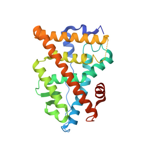
5TMW

Crystal Structure of the ER-alpha Ligand-binding Domain (Y537S) in Complex with the OBHS derivative, 4-acetamidophenyl (1S,2R,4S)-5,6-bis(4-hydroxyphenyl)-7-oxabicyclo[2.2.1]hept-5-ene-2-sulfonate


Experimental Data Snapshot

  • Method: X-RAY DIFFRACTION
  • Resolution: 2.29 Å
  • R-Value Free: 0.245 
  • R-Value Work: 0.189 
  • R-Value Observed: 0.195 

wwPDB Validation   3D Report Full Report


Ligand Structure Quality Assessment 


This is version 1.3 of the entry. See complete history


Literature

Systems Structural Biology Analysis of Ligand Effects on ER alpha Predicts Cellular Response to Environmental Estrogens and Anti-hormone Therapies.

Nwachukwu, J.C.Srinivasan, S.Bruno, N.E.Nowak, J.Wright, N.J.Minutolo, F.Rangarajan, E.S.Izard, T.Yao, X.Q.Grant, B.J.Kojetin, D.J.Elemento, O.Katzenellenbogen, J.A.Nettles, K.W.

(2017) Cell Chem Biol 24: 35-45

  • DOI: https://doi.org/10.1016/j.chembiol.2016.11.014
  • Primary Citation of Related Structures:  
    5KR9, 5KRA, 5KRC, 5KRF, 5KRH, 5KRI, 5KRJ, 5KRK, 5KRL, 5KRM, 5KRO, 5TLD, 5TLF, 5TLG, 5TLL, 5TLM, 5TLO, 5TLP, 5TLT, 5TLU, 5TLV, 5TLX, 5TLY, 5TM1, 5TM2, 5TM3, 5TM4, 5TM5, 5TM6, 5TM7, 5TM8, 5TM9, 5TML, 5TMM, 5TMO, 5TMQ, 5TMR, 5TMS, 5TMT, 5TMU, 5TMV, 5TMW, 5TMZ, 5TN1, 5TN3, 5TN4, 5TN5, 5TN6, 5TN7, 5TN8

  • PubMed Abstract: 

    Environmental estrogens and anti-hormone therapies for breast cancer have diverse tissue- and signaling-pathway-selective outcomes, but how estrogen receptor alpha (ERα) mediates this phenotypic diversity is poorly understood. We implemented a statistical approach to allow unbiased, parallel analyses of multiple crystal structures, and identified subtle perturbations of ERα structure by different synthetic and environmental estrogens. Many of these perturbations were in the sub-Å range, within the noise of the individual structures, but contributed significantly to the activities of synthetic and environmental estrogens. Combining structural perturbation data from many structures with quantitative cellular activity profiles of the ligands enabled identification of structural rules for ligand-specific allosteric signaling-predicting activity from structure. This approach provides a framework for understanding the diverse effects of environmental estrogens and for guiding iterative medicinal chemistry efforts to generate improved breast cancer therapies, an approach that can be applied to understanding other ligand-regulated allosteric signaling pathways.


  • Organizational Affiliation

    Department of Cancer Biology, The Scripps Research Institute, 130 Scripps Way, Jupiter, FL 33458, USA.


Macromolecules
Find similar proteins by:  (by identity cutoff)  |  3D Structure
Entity ID: 1
MoleculeChains Sequence LengthOrganismDetailsImage
Estrogen receptorA,
C [auth B]
257Homo sapiensMutation(s): 1 
Gene Names: ESR1ESRNR3A1
UniProt & NIH Common Fund Data Resources
Find proteins for P03372 (Homo sapiens)
Explore P03372 
Go to UniProtKB:  P03372
PHAROS:  P03372
GTEx:  ENSG00000091831 
Entity Groups  
Sequence Clusters30% Identity50% Identity70% Identity90% Identity95% Identity100% Identity
UniProt GroupP03372
Sequence Annotations
Expand
  • Reference Sequence

Find similar proteins by:  Sequence   |   3D Structure  

Entity ID: 2
MoleculeChains Sequence LengthOrganismDetailsImage
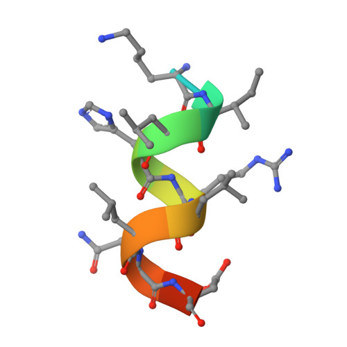
Nuclear receptor coactivator 2B [auth C],
D
13Homo sapiensMutation(s): 0 
UniProt & NIH Common Fund Data Resources
Find proteins for Q15596 (Homo sapiens)
Explore Q15596 
Go to UniProtKB:  Q15596
PHAROS:  Q15596
GTEx:  ENSG00000140396 
Entity Groups  
Sequence Clusters30% Identity50% Identity70% Identity90% Identity95% Identity100% Identity
UniProt GroupQ15596
Sequence Annotations
Expand
  • Reference Sequence
Small Molecules
Ligands 1 Unique
IDChains Name / Formula / InChI Key2D Diagram3D Interactions
7FP
Query on 7FP

Download Ideal Coordinates CCD File 
E [auth A],
F [auth B]
4-(acetylamino)phenyl (1S,2R,4S)-5,6-bis(4-hydroxyphenyl)-7-oxabicyclo[2.2.1]hept-5-ene-2-sulfonate
C26 H23 N O7 S
MNNBNPWDCGOXMP-PPJWLVRDSA-N
Experimental Data & Validation

Experimental Data

  • Method: X-RAY DIFFRACTION
  • Resolution: 2.29 Å
  • R-Value Free: 0.245 
  • R-Value Work: 0.189 
  • R-Value Observed: 0.195 
  • Space Group: P 1 21 1
Unit Cell:
Length ( Å )Angle ( ˚ )
a = 56.152α = 90
b = 79.188β = 110.03
c = 58.485γ = 90
Software Package:
Software NamePurpose
HKL-2000data scaling
PHENIXrefinement
PDB_EXTRACTdata extraction
HKL-2000data reduction
PHENIXphasing
DENZOdata reduction
SCALEPACKdata scaling

Structure Validation

View Full Validation Report


Ligand Structure Quality Assessment 


Entry History 

Revision History  (Full details and data files)

  • Version 1.0: 2017-01-18
    Type: Initial release
  • Version 1.1: 2017-02-01
    Changes: Database references
  • Version 1.2: 2017-11-22
    Changes: Refinement description
  • Version 1.3: 2024-03-06
    Changes: Data collection, Database references