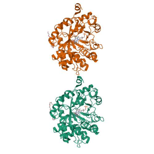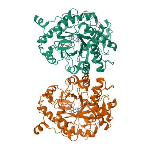
Saccharomyces cerevisiae OYE 3 soaked with p-hydroxybenzaldehyde
Stewart, J., Powell III, R.W.To be published.
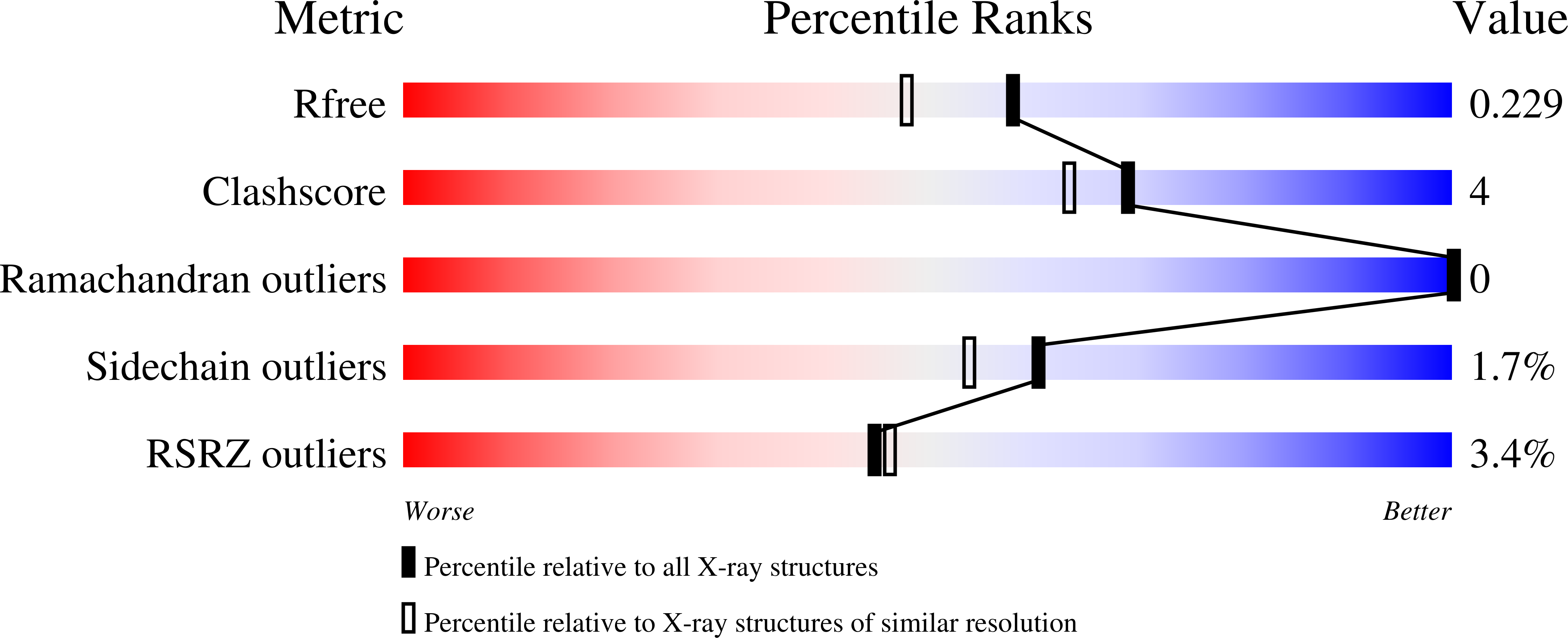
Experimental Data Snapshot
Entity ID: 1 | |||||
|---|---|---|---|---|---|
| Molecule | Chains | Sequence Length | Organism | Details | Image |
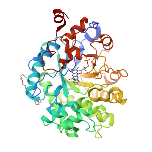
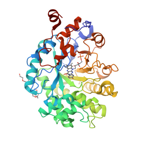
| NADPH dehydrogenase 3 | 400 | Saccharomyces cerevisiae S288C | Mutation(s): 0 Gene Names: OYE3, YPL171C, P2291 EC: 1.6.99.1 | 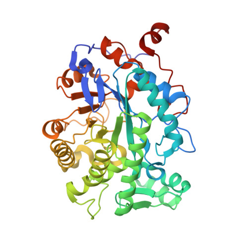 | |
UniProt | |||||
Find proteins for P41816 (Saccharomyces cerevisiae (strain ATCC 204508 / S288c)) Explore P41816 Go to UniProtKB: P41816 | |||||
Entity Groups | |||||
| Sequence Clusters | 30% Identity50% Identity70% Identity90% Identity95% Identity100% Identity | ||||
| UniProt Group | P41816 | ||||
Sequence AnnotationsExpand | |||||
| |||||
| Ligands 4 Unique | |||||
|---|---|---|---|---|---|
| ID | Chains | Name / Formula / InChI Key | 2D Diagram | 3D Interactions | |
| FMN Query on FMN | C [auth A], H [auth B] | FLAVIN MONONUCLEOTIDE C17 H21 N4 O9 P FVTCRASFADXXNN-SCRDCRAPSA-N |  | ||
| TOE Query on TOE | F [auth A], G [auth A], K [auth B], L [auth B] | 2-[2-(2-METHOXY-ETHOXY)-ETHOXY]-ETHOXYL C7 H16 O4 JLGLQAWTXXGVEM-UHFFFAOYSA-N |  | ||
| HBA Query on HBA | D [auth A], I [auth B] | P-HYDROXYBENZALDEHYDE C7 H6 O2 RGHHSNMVTDWUBI-UHFFFAOYSA-N |  | ||
| NA Query on NA | E [auth A], J [auth B] | SODIUM ION Na FKNQFGJONOIPTF-UHFFFAOYSA-N |  | ||
| Length ( Å ) | Angle ( ˚ ) |
|---|---|
| a = 61.533 | α = 90 |
| b = 106.421 | β = 90 |
| c = 141.01 | γ = 90 |
| Software Name | Purpose |
|---|---|
| PHENIX | refinement |
| XDS | data reduction |
| XDS | data scaling |
| PHENIX | phasing |
| Funding Organization | Location | Grant Number |
|---|---|---|
| National Science Foundation (NSF, United States) | United States | CHE-1111791 |
RCSB PDB is hosted by

RCSB PDB is a member of the