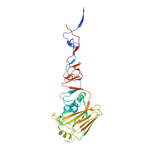
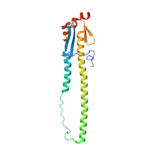
5VJK

Crystal structure of H7 hemagglutinin mutant (V186K, K193T, G228S) from the influenza virus A/Shanghai/2/2013 (H7N9)


Experimental Data Snapshot

  • Method: X-RAY DIFFRACTION
  • Resolution: 2.59 Å
  • R-Value Free: 0.251 
  • R-Value Work: 0.203 
  • R-Value Observed: 0.205 

Starting Model: experimental
View more details

wwPDB Validation   3D Report Full Report


Ligand Structure Quality Assessment 


This is version 2.1 of the entry. See complete history


Literature

Three mutations switch H7N9 influenza to human-type receptor specificity.

de Vries, R.P.Peng, W.Grant, O.C.Thompson, A.J.Zhu, X.Bouwman, K.M.de la Pena, A.T.T.van Breemen, M.J.Ambepitiya Wickramasinghe, I.N.de Haan, C.A.M.Yu, W.McBride, R.Sanders, R.W.Woods, R.J.Verheije, M.H.Wilson, I.A.Paulson, J.C.

(2017) PLoS Pathog 13: e1006390-e1006390

  • DOI: https://doi.org/10.1371/journal.ppat.1006390
  • Primary Citation of Related Structures:  
    5VJK, 5VJL, 5VJM

  • PubMed Abstract: 

    The avian H7N9 influenza outbreak in 2013 resulted from an unprecedented incidence of influenza transmission to humans from infected poultry. The majority of human H7N9 isolates contained a hemagglutinin (HA) mutation (Q226L) that has previously been associated with a switch in receptor specificity from avian-type (NeuAcα2-3Gal) to human-type (NeuAcα2-6Gal), as documented for the avian progenitors of the 1957 (H2N2) and 1968 (H3N2) human influenza pandemic viruses. While this raised concern that the H7N9 virus was adapting to humans, the mutation was not sufficient to switch the receptor specificity of H7N9, and has not resulted in sustained transmission in humans. To determine if the H7 HA was capable of acquiring human-type receptor specificity, we conducted mutation analyses. Remarkably, three amino acid mutations conferred a switch in specificity for human-type receptors that resembled the specificity of the 2009 human H1 pandemic virus, and promoted binding to human trachea epithelial cells.


  • Organizational Affiliation

    Departments of Molecular Medicine, & Immunology and Microbiology, The Scripps Research Institute, La Jolla, CA, United States of America.


Macromolecules
Find similar proteins by:  (by identity cutoff)  |  3D Structure
Entity ID: 1
MoleculeChains Sequence LengthOrganismDetailsImage
Hemagglutinin HA1325Influenza A virus (A/Shanghai/02/2013(H7N9))Mutation(s): 3 
Gene Names: HA
UniProt
Find proteins for R4NN21 (Influenza A virus)
Explore R4NN21 
Go to UniProtKB:  R4NN21
Entity Groups  
Sequence Clusters30% Identity50% Identity70% Identity90% Identity95% Identity100% Identity
UniProt GroupR4NN21
Glycosylation
Glycosylation Sites: 2
Sequence Annotations
Expand
  • Reference Sequence
Find similar proteins by:  (by identity cutoff)  |  3D Structure
Entity ID: 2
MoleculeChains Sequence LengthOrganismDetailsImage
Hemagglutinin HA2183Influenza A virus (A/Shanghai/02/2013(H7N9))Mutation(s): 0 
Gene Names: HA
UniProt
Find proteins for R4NN21 (Influenza A virus)
Explore R4NN21 
Go to UniProtKB:  R4NN21
Entity Groups  
Sequence Clusters30% Identity50% Identity70% Identity90% Identity95% Identity100% Identity
UniProt GroupR4NN21
Glycosylation
Glycosylation Sites: 1
Sequence Annotations
Expand
  • Reference Sequence
Oligosaccharides

Help

Entity ID: 3
MoleculeChains Length2D Diagram Glycosylation3D Interactions
beta-D-mannopyranose-(1-4)-2-acetamido-2-deoxy-beta-D-glucopyranose-(1-4)-2-acetamido-2-deoxy-beta-D-glucopyranose
C
3N-Glycosylation
Glycosylation Resources
GlyTouCan:  G15407YE
GlyCosmos:  G15407YE
GlyGen:  G15407YE
Experimental Data & Validation

Experimental Data

  • Method: X-RAY DIFFRACTION
  • Resolution: 2.59 Å
  • R-Value Free: 0.251 
  • R-Value Work: 0.203 
  • R-Value Observed: 0.205 
  • Space Group: H 3 2
Unit Cell:
Length ( Å )Angle ( ˚ )
a = 115.44α = 90
b = 115.44β = 90
c = 294.871γ = 120
Software Package:
Software NamePurpose
PHENIXrefinement
HKL-2000data reduction
HKL-2000data scaling
PHASERphasing

Structure Validation

View Full Validation Report


Ligand Structure Quality Assessment 


Entry History 

Deposition Data

Revision History  (Full details and data files)

  • Version 1.0: 2017-05-24
    Type: Initial release
  • Version 1.1: 2017-05-31
    Changes: Data collection
  • Version 1.2: 2017-06-28
    Changes: Database references
  • Version 2.0: 2020-07-29
    Type: Remediation
    Reason: Carbohydrate remediation
    Changes: Atomic model, Data collection, Derived calculations, Structure summary
  • Version 2.1: 2023-10-04
    Changes: Data collection, Database references, Refinement description, Structure summary